 Login / Register


- Clone
- M1/70 (See other available formats)
- Regulatory Status
- RUO
- Other Names
- αM integrin, Mac-1, Mo1, CR3, Ly-40, C3biR, ITGAM
- Isotype
- Rat IgG2b, κ
- Ave. Rating
- Submit a Review
- Product Citations
- publications

-

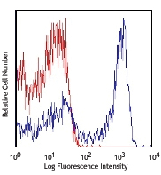
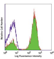
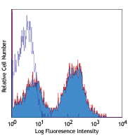
C57BL/6 mouse bone marrow cells were stained with CD11b (clone M1/70) Alexa Fluor® 488 (filled histogram) or rat IgG2b Alexa Fluor® 488 isotype control (open histogram) (gated on total cell population). -

C57BL/6 mouse frozen spleen section was fixed with 4% paraformaldehyde (PFA) for ten minutes at room temperature and blocked with 5% FBS for 30 minutes at room temperature. Then the section was stained with 2.5 µg/ml of CD11b (clone M1/70) Alexa Fluor® 488 (green), and co-stained with 5 µg/ml of CD3 (clone 17A2) Alexa Fluor® 647 (cyan) and 5 µg/ml of CD45R/B220 (clone RA3-6B2) Alexa Fluor® 594 (red) overnight at 4°C. The image was captured with a 10X objective. -

Paraformaldehyde-fixed (1%), 500 µm-thick mouse spleen section was processed according to the Ce3D™ Tissue Clearing Kit protocol (Cat. No. 427701). The section was costained with anti-mouse/human CD11b Antibody (clone M1/70) Alexa Fluor® 488 at 5 µg/mL (green), and anti-mouse IgD Antibody (clone 11-26c.2a) Alexa Fluor® 594 at 5 µg/mL (magenta). The section was then optically cleared and mounted in a sample chamber. The image was captured with a 10X objective using Zeiss 780 confocal microscope and processed by Imaris image analysis software.
Watch the video. -

Confocal image of C57BL/6 mouse liver sample acquired using the IBEX method of highly multiplexed antibody-based imaging: CD4 (yellow) in Cycle 1, CD11c (cyan) in Cycle 4, and CD11b (purple) in Cycle 4. Tissues were prepared using ~1% (vol/vol) formaldehyde and a detergent. Following fixation, samples are immersed in 30% (wt/vol) sucrose for cryoprotection. Images are courtesy of Drs. Andrea J. Radtke and Ronald N. Germain of the Center for Advanced Tissue Imaging (CAT-I) in the National Institute of Allergy and Infectious Diseases (NIAID, NIH).
| Cat # | Size | Price | Quantity Check Availability | Save | ||
|---|---|---|---|---|---|---|
| 101219 | 25 µg | £70 | ||||
| 101217 | 100 µg | £159 | ||||
CD11b is a 170 kD glycoprotein also known as αM integrin, Mac-1 α subunit, Mol, CR3, and Ly-40. CD11b is a member of the integrin family, primarily expressed on granulocytes, monocytes/macrophages, dendritic cells, NK cells, and subsets of T and B cells. CD11b non-covalently associates with CD18 (β2 integrin) to form Mac-1. Mac-1 plays an important role in cell-cell interaction by binding its ligands ICAM-1 (CD54), ICAM-2 (CD102), ICAM-4 (CD242), iC3b, and fibrinogen.
Product DetailsProduct Details
- Verified Reactivity
- Mouse, Human, Cynomolgus, Rhesus
- Reported Reactivity
- Chimpanzee, Baboon, Rabbit
- Antibody Type
- Monoclonal
- Host Species
- Rat
- Immunogen
- C57BL/10 splenocytes
- Formulation
- Phosphate-buffered solution, pH 7.2, containing 0.09% sodium azide.
- Preparation
- The antibody was purified by affinity chromatography and conjugated with Alexa Fluor® 488 under optimal conditions.
- Concentration
- 0.5 mg/ml
- Storage & Handling
- The antibody solution should be stored undiluted between 2°C and 8°C, and protected from prolonged exposure to light. Do not freeze.
- Application
-
FC - Quality tested
IHC-F, 3D IHC - Verified
SB - Community verified
SB - Reported in the literature, not verified in house - Recommended Usage
-
Each lot of this antibody is quality control tested by immunofluorescent staining with flow cytometric analysis. For flow cytometric staining, the suggested use of this reagent is ≤ 0.25 µg per 106 cells in 100 µl volume. For immunohistochemical staining on frozen tissue sections, the suggested use of this reagent is 2.5 - 10 µg per ml. For 3D immunohistochemistry on formalin-fixed tissues, a concentration of 5.0 µg/mL is suggested. It is recommended that the reagent be titrated for optimal performance for each application.
* Alexa Fluor® 488 has a maximum emission of 519 nm when it is excited at 488 nm.
Alexa Fluor® and Pacific Blue™ are trademarks of Life Technologies Corporation.
View full statement regarding label licenses - Excitation Laser
-
Blue Laser (488 nm)
- Application Notes
-
Clone M1/70 has been verified for immunocytochemistry (ICC) and frozen immunohistochemistry (IHC-F).
Additional reported applications (for relevant formats of this clone) include: immunoprecipitation1,4, in vitro blocking3,9,12, depletion2,8, immunofluorescence microscopy6,7,10, immunohistochemistry of acetone-fixed frozen sections5,11-13, and spatial biology (IBEX)35,36. For in vivo studies or highly sensitive assays, we recommend Ultra-LEAF™ purified antibody (Endotoxin < 0.01 EU/µg, Azide-Free, 0.2 µm filtered) (Cat. No. 101248). - Additional Product Notes
-
For use in spatial biology, this antibody has been demonstrated for use in immunohistochemistry using IBEX (Reported in the literature, not verified in house) and the NanoString GeoMx® Digital Spatial Profiler.
IBEX: Iterative Bleaching Extended multi-pleXity (IBEX) is a fluorescent imaging technique capable of highly-multiplexed spatial analysis. The method relies on cyclical bleaching of panels of fluorescent antibodies in order to image and analyze many markers over multiple cycles of staining, imaging, and, bleaching. It is a community-developed open-access method developed by the Center for Advanced Tissue Imaging (CAT-I) in the National Institute of Allergy and Infectious Diseases (NIAID, NIH).
NanoString GeoMx®: This product has been verified for IHC-F (Immunohistochemistry - frozen tissue sections) and IHC-P (Immunohistochemistry - formalin-fixed paraffin-embedded tissues) on the NanoString GeoMx® Digital Spatial Profiler. The GeoMx® enables researchers to perform spatial analysis of protein and RNA targets in FFPE and fresh frozen human and mouse samples. For more information about our spatial biology products and the GeoMx® platform, please visit our spatial biology page. -
Application References
(PubMed link indicates BioLegend citation) -
- Springer T, et al. 1978. Eur. J. Immunol. 8:539. (IP)
- Ault K and Springer T. 1981. J. Immunol. 126:359. (Deplete)
- Springer TA, et al. 1982. Immunol. Rev. 68:171. (Block)
- Ho MK and Springer TA. 1983. J. Biol. Chem. 258:2766. (IP)
- Flotte TJ, et al. 1983. Am. J. Pathol. 111:112. (IHC)
- Noel GJ, et al. 1990. J. Clin. Invest. 85:208. (IF)
- Allen LA and Aderem A. 1996. J. Exp. Med. 184:627 (IF)
- D'Amico A and Wu L. 2003. J. Exp. Med. 198:293. (Deplete)
- Brickson SJ, et al. 2003. Appl Physiol. 95:969. (Block)
- Clatworthy MR and Smith KG. 2004. J. Exp. Med. 199:717. (IF)
- Hata H, et al. 2004. J. Clin. Invest. 114:582. (IHC)
- Zhang Y, et al. 2002. J. Immunol. 168:3088. (IHC)
- Iwasaki A and Kelsall BL. 2001. J. Immunol. 166:4884 (IHC, FC)
- Tailleux L. 2003. J. Exp. Med. 197:121. (Block, FC)
- Olver S, et al. 2006. Cancer Research 66:571. (FC)
- Tan SL, et al. 2006. J. Immunol. 176:2872. (FC) PubMed
- Ponomarev ED, et al. 2006. J. Immunol. 176:1402. (FC)
- Dzhagalov I, et al. 2007. Blood 109:1620. (FC)
- Fazilleau N, et al. 2007. Nature Immunol. 8:753.
- Rasmussen JW, et al. 2006. Infect. Immun.74:6590. PubMed
- Napimoga MH, et al. 2008. J. Immunol. 180:609. PubMed
- Elqaraz-Carmon V, et al. 2008. J. Lipid. Res. 49:1894. PubMed
- Kim DD, et al. 2008. Blood 112:1109. PubMed
- Guo Y, et al. 2008. Blood 112:480. PubMed
- Norian LA, et al. 2009. Cancer Res. 69:3086. (FC) PubMed
- Baumgartner CK, et al. 2010. J. Immunol. 184:573. PubMed
- Charles N, et al. 2010. Nat. Med. 16:701. (FC) PubMed
- Whiteland J, et al. 1995. J. Histochem. Cytochem. 43:313. (IHC)
- Weber GF, et al. 2014. J Exp Med. 211:1243. PubMed
- Ashok A, et al. 2015. Toxicol Sci. 143:64. PubMed
- Price PJ, et al. 2015. J Immunol. 194:1164. PubMed
- Doni A, et al. 2015. J Exp Med. 212:905. PubMed
- Ferreira R, et al. 2016. J Infect Dis. 213: 669 - 673. PubMed
- Peterson VM, et al. 2017. Nat. Biotechnol. 35:936. (PG)
- Radtke AJ, et al. 2020. Proc Natl Acad Sci U S A. 117:33455-65. (SB) PubMed
- Radtke AJ, et al. 2022. Nat Protoc. 17:378-401. (SB) PubMed
- Product Citations
-
- RRID
-
AB_493545 (BioLegend Cat. No. 101219)
AB_389305 (BioLegend Cat. No. 101217)
Antigen Details
- Structure
- Integrin family, associates with integrin β2 (CD18), 170 kD
- Distribution
-
Granulocytes, monocytes/macrophages, dendritic cells, NK cells, subsets of T and B cells
- Function
- Adhesion, chemotaxis
- Ligand/Receptor
- ICAM-1 (CD54), ICAM-2 (CD102), ICAM-4 (CD242), iC3b, fibrinogen
- Cell Type
- B cells, Dendritic cells, Granulocytes, Macrophages, Monocytes, Neutrophils, NK cells, T cells, Tregs
- Biology Area
- Cell Adhesion, Cell Biology, Costimulatory Molecules, Immunology, Innate Immunity, Neuroscience, Neuroscience Cell Markers
- Molecular Family
- Adhesion Molecules, CD Molecules
- Antigen References
-
1. Barclay A, et al. 1997. The Leukocyte Antigen FactsBook Academic Press.
2. Springer TA. 1994. Cell 76:301.
3. Coxon A, et al. 1996. Immunity 5:653. - Gene ID
- 16409 View all products for this Gene ID 3684 View all products for this Gene ID
- UniProt
- View information about CD11b on UniProt.org
Related Pages & Pathways
Pathways


Related FAQs
- If an antibody clone has been previously successfully used in IBEX in one fluorescent format, will other antibody formats work as well?
-
It’s likely that other fluorophore conjugates to the same antibody clone will also be compatible with IBEX using the same sample fixation procedure. Ultimately a directly conjugated antibody’s utility in fluorescent imaging and IBEX may be specific to the sample and microscope being used in the experiment. Some antibody clone conjugates may perform better than others due to performance differences in non-specific binding, fluorophore brightness, and other biochemical properties unique to that conjugate.
- Will antibodies my lab is already using for fluorescent or chromogenic IHC work in IBEX?
-
Fundamentally, IBEX as a technique that works much in the same way as single antibody panels or single marker IF/IHC. If you’re already successfully using an antibody clone on a sample of interest, it is likely that clone will have utility in IBEX. It is expected some optimization and testing of different antibody fluorophore conjugates will be required to find a suitable format; however, legacy microscopy techniques like chromogenic IHC on fixed or frozen tissue is an excellent place to start looking for useful antibodies.
- Are other fluorophores compatible with IBEX?
-
Over 18 fluorescent formats have been screened for use in IBEX, however, it is likely that other fluorophores are able to be rapidly bleached in IBEX. If a fluorophore format is already suitable for your imaging platform it can be tested for compatibility in IBEX.
- The same antibody works in one tissue type but not another. What is happening?
-
Differences in tissue properties may impact both the ability of an antibody to bind its target specifically and impact the ability of a specific fluorophore conjugate to overcome the background fluorescent signal in a given tissue. Secondary stains, as well as testing multiple fluorescent conjugates of the same clone, may help to troubleshoot challenging targets or tissues. Using a reference control tissue may also give confidence in the specificity of your staining.
- How can I be sure the staining I’m seeing in my tissue is real?
-
In general, best practices for validating an antibody in traditional chromogenic or fluorescent IHC are applicable to IBEX. Please reference the Nature Methods review on antibody based multiplexed imaging for resources on validating antibodies for IBEX.
Other Formats
View All CD11b Reagents Request Custom ConjugationCustomers Also Purchased
Compare Data Across All Formats
This data display is provided for general comparisons between formats.
Your actual data may vary due to variations in samples, target cells, instruments and their settings, staining conditions, and other factors.
If you need assistance with selecting the best format contact our expert technical support team.
-
APC anti-mouse/human CD11b

C57BL/6 mouse bone marrow cells were stained with CD11b (clo... -
Biotin anti-mouse/human CD11b

C57BL/6 mouse bone marrow cells were stained with biotinylat... -
FITC anti-mouse/human CD11b

C57BL/6 mouse bone marrow cells were stained with CD11b (clo... 
-
PE anti-mouse/human CD11b

C57BL/6 mouse bone marrow cells were stained with CD11b (clo... 
Fixed whole mount mouse spleen was stained with Alexa Fluor®... -
PE/Cyanine5 anti-mouse/human CD11b

C57BL/6 mouse bone marrow cells stained with CD11b (clone M1... -
Purified anti-mouse/human CD11b

C57BL/6 mouse bone marrow cells were stained with purified C... 
Fresh, frozen mouse spleen was stained with purified CD11b c... -
PE/Cyanine7 anti-mouse/human CD11b

C57BL/6 mouse bone marrow cells were stained with CD11b (clo... -
Alexa Fluor® 488 anti-mouse/human CD11b

C57BL/6 mouse bone marrow cells were stained with CD11b (clo... 
C57BL/6 mouse frozen spleen section was fixed with 4% parafo... 
Paraformaldehyde-fixed (1%), 500 µm-thick mouse spleen secti... 
Confocal image of C57BL/6 mouse liver sample acquired using ... -
Alexa Fluor® 647 anti-mouse/human CD11b

C57BL/6 mouse bone marrow cells were stained with CD11b (clo... 
Paraformaldehyde-fixed (4%), 500 µm-thick mouse spleen secti... -
Alexa Fluor® 700 anti-mouse/human CD11b

C57BL/6 mouse bone marrow cells were stained with CD11b (clo... -
Pacific Blue™ anti-mouse/human CD11b

C57BL/6 mouse bone marrow cells were stained with CD11b (clo... -
APC/Cyanine7 anti-mouse/human CD11b

C57BL/6 mouse bone marrow stained with M1/70 APC/Cyanine7 -
PerCP/Cyanine5.5 anti-mouse/human CD11b

C57BL/6 splenocytes were blocked with TruStain FcX™ (a... -
PerCP anti-mouse/human CD11b

C57BL/6 mouse bone marrow cells were stained with CD11b (clo... -
Brilliant Violet 421™ anti-mouse/human CD11b

C57BL/6 mouse bone marrow cells were stained with CD11b (clo... 
BL/6 mouse lymph nodes, fixed O/N in PLP, blocked with 10% r... -
Brilliant Violet 570™ anti-mouse/human CD11b

C57BL/6 mouse bone marrow cells were stained with CD11b (clo... -
Brilliant Violet 605™ anti-mouse/human CD11b

C57BL/6 mouse bone marrow cells were stained with CD11b (clo... -
Brilliant Violet 650™ anti-mouse/human CD11b

C57BL/6 mouse bone marrow cells were stained with CD11b (clo... -
Brilliant Violet 711™ anti-mouse/human CD11b

C57BL/6 mouse bone marrow cells were stained with CD11b (clo... -
Brilliant Violet 785™ anti-mouse/human CD11b

C57BL/6 mouse bone marrow cells were stained with CD11b (clo... -
Brilliant Violet 510™ anti-mouse/human CD11b

C57BL/6 mouse bone marrow cells were stained with CD11b (clo... 
C57 mouse bone marrow cells were fixed with 2% paraformaldeh... -
Ultra-LEAF™ Purified anti-mouse/human CD11b

C57BL/6 mouse bone marrow cells were stained with Ultra-LEAF... -
Purified anti-mouse/human CD11b (Maxpar® Ready)

Mouse splenocytes stained with 172Yb-anti-CD11b (M1/70) and ... -
Alexa Fluor® 594 anti-mouse/human CD11b

C57BL/6 mouse frozen spleen section was fixed with 4% parafo... 
C57BL/6 mouse bone marrow cells were stained with CD11b (clo... -
PE/Dazzle™ 594 anti-mouse/human CD11b

C57BL/6 mouse bone marrow cells were stained with CD11b (clo... -
APC/Fire™ 750 anti-mouse/human CD11b

C57BL/6 splenocytes were blocked with TruStain fcX™ (a... 
-
TotalSeq™-A0014 anti-mouse/human CD11b
-
Brilliant Violet 750™ anti-mouse/human CD11b

C57BL/6 mouse bone marrow cells were stained with Ly-6G/Ly-6... -
TotalSeq™-B0014 anti-mouse/human CD11b
-
TotalSeq™-C0014 anti-mouse/human CD11b
-
Spark NIR™ 685 anti-mouse/human CD11b

C57BL/6 splenocytes were stained with Ly-6G/Ly-6C (Gr-1) Pac... -
PE/Fire™ 640 anti-mouse/human CD11b

C57BL/6 mouse bone marrow cells were stained with anti-mouse... -
Spark YG™ 593 anti-mouse/human CD11b

C57BL/6 mouse bone marrow cells were stained with Ly-6G/Ly-6... -
Spark YG™ 570 anti-mouse/human CD11b

C57BL/6 mouse frozen spleen section was fixed with 4% parafo... -
PE/Fire™ 810 anti-mouse/human CD11b

C57BL/6 mouse bone marrow cells were stained with anti-mouse... -
APC/Fire™ 810 anti-mouse/human CD11b Antibody

C57BL/6 mouse bone marrow cells were stained with anti-mouse... -
Spark Blue™ 550 anti-mouse/human CD11b

C57BL/6 mouse bone marrow cells were stained with anti-mouse... -
Spark UV™ 387 anti-mouse/human CD11b

C57BL/6 bone marrow cells were stained with anti-mouse Ly-6G... -
PerCP/Fire™ 806 anti-mouse/human CD11b

C57BL/6 mouse bone marrow cells were stained with anti-mouse... -
PerCP/Fire™ 780 anti-mouse/human CD11b

C57BL/6 mouse bone marrow cells were stained with anti-mouse... -
Spark Blue™ 574 anti-mouse/human CD11b (Flexi-Fluor™)
-
KIRAVIA Blue 520™ anti-mouse/human CD11b

C57BL/6 mouse bone marrow cells were stained with anti-mouse... -
PE/Fire™ 744 anti-mouse/human CD11b

C57BL/6 mouse bone marrow cells were stained with anti-mouse... -
Spark PLUS UV™ 395 anti-mouse/human CD11b

C57BL/6 mouse splenocytes were stained with anti-mouse Ly-6G...


Follow Us