


- Clone
- M380B (See other available formats)
- Regulatory Status
- RUO
- Isotype
- Mouse IgG1
- Ave. Rating
- Submit a Review
- Product Citations
- publications

-

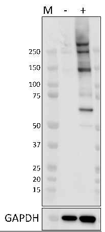
Whole cell lysates (15 μg) from untreated (-) or Calyculin A treated (+) (50 nM for 30 min) HeLa cells were resolved by 4-12% Bis-Tris gel electrophoresis, transferred to a PVDF membrane and probed with 1.0 μg/mL (1:500 dilution) purified anti-Phosphoserine antibody (clone M380B) overnight at 4°C. Proteins were visualized by chemiluminescence detection using HRP goat anti-mouse IgG antibody (Cat. No. 405306) at a 1:3000 dilution. Direct-Blot™ HRP anti-GAPDH antibody (Cat. No. 607904) was used as a loading control at a 1:50000 dilution (lower). Lane M: Molecular weight marker. -

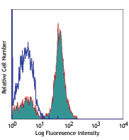
HeLa cells were fixed with 4% paraformaldehyde for 10 minutes, permeabilized with 0.5% Triton-X for 10 minutes, and blocked with 5% FBS for 30 minutes. Cells were then intracellularly stained with a 5.0 µg/mL (1:100 dilution) (panel A) purified anti-Phosphoserine antibody (clone M380B) overnight at 4°C, followed by incubation with Alexa Fluor® 594 goat anti-mouse IgG antibody (Cat. No. 405326) at 2.5 µg/mL. To demonstrate phospho-specificity of M380B, fixed and permeabilized HeLa cells were pre-treated with lambda protein phosphatase as a control prior to staining with M380B (panel B). Nuclei were counterstained with DAPI, and the image was captured with a 60X objective. -

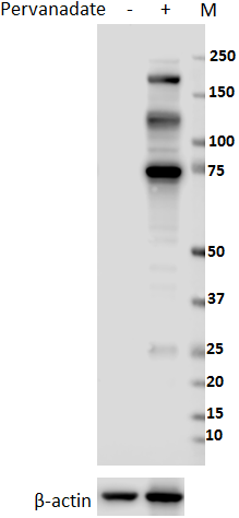
Whole cell extracts (250 µg total protein) prepared from HeLa cells untreated (-) or treated (+) with Calyculin A (50 nM, 30 min) were immunoprecipitated overnight with 2.5 µg purified mouse IgG1, κ isotype ctrl antibody (Cat. No. 401401) or purified anti-Phosphoserine antibody (clone M380B). The resulting IP fractions and whole cell extract input (10%) were resolved by 4-12% Bis-Tris gel electrophoresis, transferred to a PVDF membrane and probed with purified anti-Phosphoserine antibody (clone M380B). Lane M: Molecular weight marker.
| Cat # | Size | Price | Save |
|---|---|---|---|
| 944101 | 25 µg | ¥25,960 | |
| 944102 | 100 µg | ¥64,460 |
Phosphorylation of specific serine residues is a post-translational modification event critical for regulating the activity of many proteins. Protein phosphorylation events can alter protein function through a variety of molecular mechanisms, ranging from conformational changes that can stimulate or inhibit protein function to facilitating protein-protein interactions with binding partners. Antibodies that can detect phosphoserine residues are valuable tools for identifying novel phospho-sites and characterizing changes in the post-translational state of a broad range of phosphorylated proteins.
Product DetailsProduct Details
- Verified Reactivity
- Human, Mouse, Rat, All Species
- Antibody Type
- Monoclonal
- Host Species
- Mouse
- Immunogen
- Synthetic peptide containing phosphorylated serine
- Formulation
- Phosphate-buffered solution, pH 7.2, containing 0.09% sodium azide
- Preparation
- The antibody was purified by affinity chromatography.
- Concentration
- 0.5 mg/mL
- Storage & Handling
- The antibody solution should be stored undiluted between 2°C and 8°C.
- Application
-
WB - Quality tested
ICC, IP - Verified - Recommended Usage
-
Each lot of this antibody is quality control tested by western blotting. For western blotting, the suggested use of this reagent is 1.0 µg/mL. For immunocytochemistry, a concentration range of 1.0 - 5.0 μg/mL is recommended. For immunoprecipitation, the suggested use of this reagent is 2.5 µg/test. It is recommended that the reagent be titrated for optimal performance for each application.
- RRID
-
AB_2890869 (BioLegend Cat. No. 944101)
AB_2890869 (BioLegend Cat. No. 944102)
Antigen Details
- Function
- Cell signaling
- Biology Area
- Cell Biology, Immunology, Signal Transduction
- Molecular Family
- Phospho-Proteins, Protein Kinases/Phosphatase
- Antigen References
-
1. Rogerson D. et. al. 2015. Nat. Chem. Biol. 11:496-503.
2. Steinfield J. et. al. 2014. ACS Chem. Biol. 9(5):1104-1112. - Gene ID
- NA
- UniProt
- View information about Phosphoserine on UniProt.org

Related FAQs
Other Formats
View All Phosphoserine Reagents Request Custom Conjugation| Description | Clone | Applications |
|---|---|---|
| Purified anti-Phosphoserine | M380B | WB,ICC,IP |
Customers Also Purchased
Compare Data Across All Formats
This data display is provided for general comparisons between formats.
Your actual data may vary due to variations in samples, target cells, instruments and their settings, staining conditions, and other factors.
If you need assistance with selecting the best format contact our expert technical support team.


Follow Us