 Login / Register


- Clone
- MR1 (See other available formats)
- Regulatory Status
- RUO
- Other Names
- CD40 ligand (CD40L), gp39, T-BAM, TRAP, Ly-62, TNFSF5
- Isotype
- Armenian Hamster IgG
- Ave. Rating
- Submit a Review
- Product Citations
- publications

-

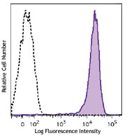
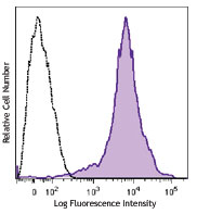
Enriched C57BL/6 mouse splenic T cells were stimulated with PMA+ionomycin for 6 hours and then stained with CD154 (clone MR1) PE/Cyanine7 (filled histogram) or Armenian hamster IgG PE/Cyanine7 (open histogram).
| Cat # | Size | Price | Quantity Check Availability | Save | ||
|---|---|---|---|---|---|---|
| 106511 | 25 µg | £85 | ||||
| 106512 | 100 µg | £197 | ||||
CD154 is a 39 kD TNF superfamily member also known as CD40 ligand, gp39, T-BAM, TRAP, and Ly-62. CD154 is an accessory molecule expressed predominantly on activated CD4+ lymphocytes that bind CD40. CD154 plays an important role in T-B cell costimulation. The MR1 antibody has been reported to inhibit the activation of T and B lymphocytes in vitro and antigen-specific lymphocyte responses in vivo.
Product DetailsProduct Details
- Verified Reactivity
- Mouse
- Antibody Type
- Monoclonal
- Host Species
- Armenian Hamster
- Immunogen
- Activated mouse Th1 clone D1.6
- Formulation
- Phosphate-buffered solution, pH 7.2, containing 0.09% sodium azide.
- Preparation
- The antibody was purified by affinity chromatography and conjugated with PE/Cyanine7 under optimal conditions.
- Concentration
- 0.2 mg/ml
- Storage & Handling
- The antibody solution should be stored undiluted between 2°C and 8°C, and protected from prolonged exposure to light. Do not freeze.
- Application
-
FC - Quality tested
- Recommended Usage
-
Each lot of this antibody is quality control tested by immunofluorescent staining with flow cytometric analysis. For flow cytometric staining, the suggested use of this reagent is ≤0.25 µg per million cells in 100 µl volume. It is recommended that the reagent be titrated for optimal performance for each application.
- Excitation Laser
-
Blue Laser (488 nm)
Green Laser (532 nm)/Yellow-Green Laser (561 nm)
- Application Notes
-
Additional reported applications (for the relevant formats) include: immunohistochemical staining1,2 of acetone-fixed frozen sections, and in vitro and in vivo blocking of ligand binding3-5. For most successful immunofluorescent staining results, it may be important to maximize signal over background by using a relatively bright fluorochrome-antibody conjugate (Cat. No. 106506) or by using a high sensitivity, three-layer staining technique (e.g., including a biotinylated antibody (Cat. No. 106504) or biotinylated anti-Armenian hamster IgG (Cat. No. 405501) second step, followed by SAv-PE (Cat. No. 405204). The Ultra-LEAF™ purified antibody (Endotoxin <0.1 EU/µg, Azide-Free, 0.2 µm filtered) is recommended for functional assays (Cat. Nos. 106515-106520).
- Additional Product Notes
- BioLegend is in the process of converting the name PE/Cy7 to PE/Cyanine7. The dye molecule remains the same, so you should expect the same quality and performance from our PE/Cyanine7 products. Please contact Technical Service if you have any questions.
-
Application References
(PubMed link indicates BioLegend citation) -
- Lettesjö H, et al. 2000. J. Immunol. 165:4095. (IHC)
- Dunn RJ, et al. 1997. J. Histochem. Cytochem. 45:129. (IHC)
- Noelle RJ, et al. 1992. P. Natl. Acad. Sci. USA 89:6550. (Block)
- Roy M, et al. 1995. Eur. J. Immunol. 25:596. (Block)
- Foy TM, et al. 1994. J. Exp. Med. 180:157. (Block)
- Lawson BR, et al. 2007. J. Immunol. 178:5366.
- Product Citations
-
- RRID
-
AB_2563492 (BioLegend Cat. No. 106511)
AB_2563493 (BioLegend Cat. No. 106512)
Antigen Details
- Structure
- TNF superfamily, 39 kD
- Distribution
-
Activated CD4+ T cells
- Function
- T-B cell costimulation
- Ligand/Receptor
- CD40
- Cell Type
- T cells, Tregs
- Biology Area
- Costimulatory Molecules, Immunology
- Molecular Family
- Adhesion Molecules, CD Molecules
- Antigen References
-
1. Barclay A, et al. 1997. The Leukocyte Antigen FactsBook Academic Press.
2. Noelle RJ, et al. 1992. P. Natl. Acad. Sci. USA 89:6550.
3. Bancherou J, et al. 1994. Annu. Rev. Immunol. 12:881.
4. Clark EA, et al. 1996. P. Natl. Acad. Sci. USA 83:4494. - Gene ID
- 21947 View all products for this Gene ID
- UniProt
- View information about CD154 on UniProt.org
Related Pages & Pathways
Pathways


Related FAQs
Other Formats
View All CD154 Reagents Request Custom Conjugation| Description | Clone | Applications |
|---|---|---|
| Biotin anti-mouse CD154 | MR1 | FC |
| PE anti-mouse CD154 | MR1 | FC |
| APC anti-mouse CD154 | MR1 | FC |
| PE/Cyanine7 anti-mouse CD154 | MR1 | FC |
| PerCP/Cyanine5.5 anti-mouse CD154 | MR1 | FC |
| Ultra-LEAF™ Purified anti-mouse CD154 | MR1 | FC,IHC-F,Block |
Customers Also Purchased
Compare Data Across All Formats
This data display is provided for general comparisons between formats.
Your actual data may vary due to variations in samples, target cells, instruments and their settings, staining conditions, and other factors.
If you need assistance with selecting the best format contact our expert technical support team.
-
Biotin anti-mouse CD154

PMA- and ionomycin-stimulated (6 hrs) BALB/c T cells stained... -
PE anti-mouse CD154
PMA- and ionomycin-stimulated (6 hrs) BALB/c T cells stained... -
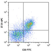
APC anti-mouse CD154

Enriched C57BL/6 mouse splenic T cells were stimulated with ... -
PE/Cyanine7 anti-mouse CD154

Enriched C57BL/6 mouse splenic T cells were stimulated with ... -
PerCP/Cyanine5.5 anti-mouse CD154

Enriched C57BL/6 mouse splenic T cells were stimulated with ... -
Ultra-LEAF™ Purified anti-mouse CD154
PMA- and ionomycin-stimulated (6 hrs) BALB/c T cells stained...


Follow Us