 Login / Register


- Clone
- WM53 (See other available formats)
- Regulatory Status
- RUO
- Workshop
- IV M-505
- Other Names
- Siglec-3, gp67, p67
- Isotype
- Mouse IgG1, κ
- Ave. Rating
- Submit a Review
- Product Citations
- publications

-

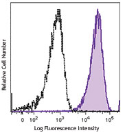
Human peripheral blood monocytes were stained with Brilliant Violet 785™ anti-human CD33 (clone WM53) (filled histogram) or Brilliant Violet 785™ mouse IgG1, κ (open histogram).
| Cat # | Size | Price | Quantity Check Availability | Save | ||
|---|---|---|---|---|---|---|
| 303427 | 25 tests | £140 | ||||
| 303428 | 100 tests | £298 | ||||
CD33 is a 67 kD type I transmembrane glycoprotein also known as Siglec-3, gp67, and p67. It is a sialoadhesion immunoglobulin superfamily member expressed on myeloid progenitors, monocytes, granulocytes, dendritic cells and mast cells. CD33 is absent on normal platelets, lymphocytes, erythrocytes and hematopoietic stem cells. CD33 functions as a sialic acid-dependent cell adhesion molecule with carbohydrate/lectin binding activity.
Product DetailsProduct Details
- Verified Reactivity
- Human
- Reported Reactivity
- Chimpanzee
- Antibody Type
- Monoclonal
- Host Species
- Mouse
- Immunogen
- Human myeloid leukaemia cells.
- Formulation
- Phosphate-buffered solution, pH 7.2, containing 0.09% sodium azide and BSA (origin USA).
- Preparation
- The antibody was purified by affinity chromatography and conjugated with Brilliant Violet 785™ under optimal conditions.
- Concentration
- Lot-specific (to obtain lot-specific concentration and expiration, please enter the lot number in our Certificate of Analysis online tool.)
- Application
-
FC - Quality tested
- Recommended Usage
-
Each lot of this antibody is quality control tested by immunofluorescent staining with flow cytometric analysis. For flow cytometric staining, the suggested use of this reagent is 5 µl per million cells in 100 µl staining volume or 5 µl per 100 µl of whole blood.
Brilliant Violet 785™ excites at 405 nm and emits at 785 nm. The bandpass filter 780/60 nm is recommended for detection, although filter optimization may be required depending on other fluorophores used. Be sure to verify that your cytometer configuration and software setup are appropriate for detecting this channel. Refer to your instrument manual or manufacturer for support. Brilliant Violet 785™ is a trademark of Sirigen Group Ltd.
Learn more about Brilliant Violet™.
This product is subject to proprietary rights of Sirigen Inc. and is made and sold under license from Sirigen Inc. The purchase of this product conveys to the buyer a non-transferable right to use the purchased product for research purposes only. This product may not be resold or incorporated in any manner into another product for resale. Any use for therapeutics or diagnostics is strictly prohibited. This product is covered by U.S. Patent(s), pending patent applications and foreign equivalents. - Excitation Laser
-
Violet Laser (405 nm)
- Application Notes
-
Additional reported applications (for the relevant formats) include: immunoprecipitation, Western blotting3, induction of cytokine production3, and immunofluorescence4. The Ultra-LEAF™ purified antibody (Endotoxin < 0.01 EU/µg, Azide-Free, 0.2 µm filtered) is recommended for functional assays (Cat. Nos. 303437 & 303438).
-
Application References
(PubMed link indicates BioLegend citation) -
- Knapp W, et al. 1989. Leucocyte Typing IV. Oxford University Press. New York.
- Favaloro E, et al. 1988. Br. J. Haematol. 69:163.
- Garnache-Ottou F, et al. 2005. Blood 105:1256. (WB)
- Pèrez-Oliva AB, et al. 2011. Glycobiology. 21:757. (epitope, FC, IF)
- Product Citations
-
- RRID
-
AB_2650887 (BioLegend Cat. No. 303427)
AB_2650888 (BioLegend Cat. No. 303428)
Antigen Details
- Structure
- Ig superfamily, sialoadhesins, type I glycoprotein, 67 kD
- Distribution
-
Myeloid progenitor, monocytes, granulocytes, dendritic cells, mast cells
- Function
- Adhesion and lectin activity
- Ligand/Receptor
- Sugar chains containing sialic acid
- Cell Type
- B cells, Dendritic cells, Granulocytes, Hematopoietic stem and progenitors, Mast cells, Monocytes, Neutrophils
- Biology Area
- Cell Biology, Immunology, Neuroinflammation, Neuroscience
- Molecular Family
- CD Molecules, Siglec Molecules
- Antigen References
-
1. Favaloro E, et al. 1988. Br. J. Haematol. 69:163.
2. Freeman S, et al. 1995. Blood 85:2005. - Gene ID
- 945 View all products for this Gene ID
- UniProt
- View information about CD33 on UniProt.org
Related Pages & Pathways
Pathways


Other Formats
View All CD33 Reagents Request Custom ConjugationCustomers Also Purchased
Compare Data Across All Formats
This data display is provided for general comparisons between formats.
Your actual data may vary due to variations in samples, target cells, instruments and their settings, staining conditions, and other factors.
If you need assistance with selecting the best format contact our expert technical support team.
-
APC anti-human CD33

Human peripheral blood lymphocytes, monocytes, and granulocy... -
PE anti-human CD33

Human peripheral blood lymphocytes, monocytes and granulocyt... -
PE/Cyanine5 anti-human CD33
-
Purified anti-human CD33

Human peripheral blood lymphocytes, monocytes, and granulocy... -
PerCP/Cyanine5.5 anti-human CD33

Human peripheral blood monocytes were stained with CD33 (clo... -
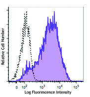
Brilliant Violet 711™ anti-human CD33

Human peripheral blood monocytes were stained with CD33 (clo... -
Brilliant Violet 421™ anti-human CD33

Human peripheral blood monocytes were stained with CD33 (clo... -
Brilliant Violet 570™ anti-human CD33

Human peripheral blood monocytes were stained with CD33 (clo... -
Purified anti-human CD33 (Maxpar® Ready)

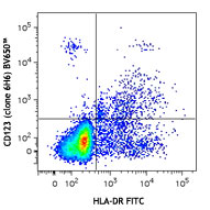
Human PBMCs stained with 166Er-anti-CD44 (BJ18) and 158Gd-an... -
Brilliant Violet 510™ anti-human CD33

Human peripheral blood monocytes were stained with CD33 (clo... -
Biotin anti-human CD33

Human peripheral blood monocytes were stained with biotinyla... -
Brilliant Violet 785™ anti-human CD33

Human peripheral blood monocytes were stained with Brilliant... -
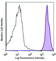
Brilliant Violet 650™ anti-human CD33

Human peripheral blood monocytes were stained with CD33 (clo... -
PE/Cyanine7 anti-human CD33

Human peripheral blood monocytes were stained with a mixture... 
Human peripheral blood lymphocytes, monocytes, and granulocy... -
PE/Dazzle™ 594 anti-human CD33

Human peripheral blood monocytes were stained with a mixture... 
Human peripheral blood lymphocytes, monocytes, and granulocy... -
APC anti-human CD33
Typical results from human peripheral blood lymphocytes, mon... -
Alexa Fluor® 700 anti-human CD33

Human peripheral blood monocytes were stained with human CD3... -
Ultra-LEAF™ Purified anti-human CD33

Human peripheral blood lymphocytes, monocytes, and granulocy... -
APC/Fire™ 750 anti-human CD33

Human peripheral blood monocytes were stained with CD33 (clo... -
PerCP/Cyanine5.5 anti-human CD33
Typical results from human peripheral blood monocytes staine... -
APC/Cyanine7 anti-human CD33

Human peripheral blood monocytes were stained with anti-huma... -
PE anti-human CD33

Typical results from human peripheral blood lymphocytes, mon... -
PE/Cyanine7 anti-human CD33

Typical results from human peripheral blood monocytes staine... -
GMP PerCP/Cyanine5.5 anti-human CD33

Typical results from human peripheral blood monocytes staine... -
Alexa Fluor® 647 anti-human CD33

Human peripheral blood monocytes were stained with anti-huma... -
GMP APC anti-human CD33

Typical results from human peripheral blood monocytes staine... -
GMP PE anti-human CD33

Typical results from human peripheral blood monocytes staine... -
PE/Fire™ 810 anti-human CD33

Human peripheral blood monocytes were stained with anti-huma... -
PE/Cyanine5 anti-human CD33

Typical results from human peripheral blood monocytes staine... -
Spark Blue™ 515 anti-human CD33

Human peripheral blood cells were surface stained with (left... -
PE/Fire™ 700 anti-human CD33

Human peripheral blood monocytes were stained with anti-huma... -
GMP PE/Cyanine5 anti-human CD33

Typical results from human peripheral blood monocytes staine... -
Spark Violet™ 423 anti-human CD33

Human peripheral blood monocytes were stained with anti-huma...


Follow Us