 Login / Register


- Clone
- M-A251 (See other available formats)
- Workshop
- IV A053
- Other Names
- IL-2 receptor α chain, Low affinity IL-2R, IL-2Rα chain
- Isotype
- Mouse IgG1, κ
- Ave. Rating
- Submit a Review
- Product Citations
- publications

-

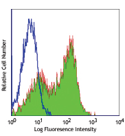
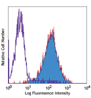
Typical results from human peripheral blood lymphocytes stained either with M-A251 PE used at 5 µL/test (filled histogram) or with an isotype control (open histogram).
| Cat # | Size | Price | Quantity Check Availability | Save | ||
|---|---|---|---|---|---|---|
| 260214 | 100 tests | 244€ | ||||
CD25 is a 55 kD type I transmembrane glycoprotein also known as low affinity IL-2 receptor α chain or Tac. It is expressed on progenitor lymphocytes, activated T and B cells, and activated monocytes/macrophages. CD25 is also expressed on a subset of non-stimulated CD4+ T cells termed T regulatory cells. Soluble CD25/IL-2Rα is produced as a consequence of lymphocyte stimulation and is found in biological fluids following inflammatory responses. CD25 associates with IL-2 receptor β (CD122) and common γ (CD132) chains to form a high affinity IL-2R complex.
Product DetailsProduct Details
- Reactivity
- Human
- Antibody Type
- Monoclonal
- Host Species
- Mouse
- Immunogen
- Human PHA-induced lymphocyte cells
- Formulation
- Phosphate-buffered solution, pH 7.2, containing 0.09% sodium azide and 0.2% (w/v) BSA (origin USA) and a stabilizer.
- Preparation
- The antibody was purified by affinity chromatography and conjugated with PE under optimal conditions.
- Concentration
- 100 µg/mL
- Storage & Handling
- The antibody solution should be stored undiluted between 2°C and 8°C, and protected from prolonged exposure to light. Do not freeze.
- Application
-
FC - Quality tested
- Recommended Usage
-
Each lot of this antibody is quality control tested by immunofluorescent staining with flow cytometric analysis. For flow cytometric staining, the suggested use of this reagent is 5 µL per million cells in 100 µL staining volume or 5 µL per 100 µL of whole blood. It is recommended that the reagent be titrated for optimal performance for each application.
- Excitation Laser
-
Blue Laser (488 nm)
Green Laser (532 nm)/Yellow-Green Laser (561 nm)
- Application Notes
-
Additional reported applications (for the relevant formats) include: immunohistochemical staining of paraformaldehyde fixed frozen sections1 and spatial biology (IBEX)2,3.
The CD25 molecule reveals three epitope regions: A, B, and C. M-A251 antibody recognizes epitope region B. Unlike other CD25 antibody clones, M-A251 can detect CD25 after fixation with paraformaldehyde. -
Application References
(PubMed link indicates BioLegend citation) - RRID
-
AB_2928019 (BioLegend Cat. No. 260214)
- Disclaimer
-
GMP RUO Flow Cytometry Antibodies. BioLegend GMP RUO fluorophore conjugated antibodies are manufactured in a dedicated GMP facility and compliant with ISO 13485:2016. For research use only. Not for use in diagnostic or therapeutic procedures. Our processes include:
- Batch-to-batch consistency
- Material traceability
- Documented procedures
- Documented employee training
- Equipment maintenance and monitoring records
- Lot-specific certificates of analysis
- Quality audits per ISO 13485:2016
- QA review of released products
Antigen Details
- Structure
- Type I transmembrane glycoprotein, 55 kD; low-affinity IL-2 receptor α chain
- Distribution
-
Activated T and B cells, monocytes/macrophages, Tregs
- Interaction
- Associates with IL-2Rβ/CD122 and IL-2Rγ/CD132 receptor chains to form a high-affinity IL-2R complex
- Ligand/Receptor
- IL-2
- Cell Type
- B cells, Macrophages, Monocytes, T cells, Tregs
- Biology Area
- Cell Biology, Immunology, Neuroscience, Neuroscience Cell Markers
- Molecular Family
- CD Molecules, Cytokine/Chemokine Receptors
- Antigen References
-
1. Knapp W, et al. 1989. Leucocyte Typing IV: White Cell Differentiation Antigens. Oxford University Press.
2. Schlossman S, et al. 1995. Leucocyte Typing V: White Cell Differentiation Antigens. Oxford University Press.
3. Barclay N, et al. 1997. The Leukocyte Antigen FactsBook. Academic Press Inc.
4. Taniguchi T and Minami Y. et al. 1993. Cell 73:5.
5. Waldmann T. 1991. J. Biol. Chem. 266:2681. - Gene ID
- 3559 View all products for this Gene ID
- UniProt
- View information about CD25 on UniProt.org
Related Pages & Pathways
Pathways


Related FAQs
- What type of PE do you use in your conjugates?
- We use R-PE in our conjugates.
Other Formats
View All CD25 Reagents Request Custom ConjugationCustomers Also Purchased
Compare Data Across All Formats
This data display is provided for general comparisons between formats.
Your actual data may vary due to variations in samples, target cells, instruments and their settings, staining conditions, and other factors.
If you need assistance with selecting the best format contact our expert technical support team.
-
APC/Cyanine7 anti-human CD25

PHA-stimulated (3 days) human peripheral blood lymphocytes w... -
Purified anti-human CD25

PHA-stimulated (3 day) human peripheral blood lymphocytes we... -
PE anti-human CD25

PHA-stimulated (3 day) human peripheral blood lymphocytes we... -
FITC anti-human CD25

PHA-stimulated (3 day) human peripheral blood lymphocytes we... -
PE/Cyanine7 anti-human CD25

PHA-stimulated (3 days) human peripheral blood lymphocytes w... -
APC anti-human CD25

PHA-stimulated (3 days) human peripheral blood lymphocytes w... -
PerCP/Cyanine5.5 anti-human CD25

PHA-stimulated (3 days) human peripheral blood lymphocytes w... -
Brilliant Violet 421™ anti-human CD25

PHA-stimulated (3 days) human peripheral blood lymphocytes w... -
Alexa Fluor® 488 anti-human CD25

PHA-stimulated (3 days) human peripheral blood lymphocytes w... -
Alexa Fluor® 700 anti-human CD25

PHA-stimulated (3 days) human peripheral blood lymphocytes w... -
Brilliant Violet 510™ anti-human CD25

PHA-stimulated (3 days) human peripheral blood lymphocytes w... -
PE/Dazzle™ 594 anti-human CD25

PHA-stimulated (3 days) human peripheral blood lymphocytes w... -
Biotin anti-human CD25

PHA-stimulated (3 days) human peripheral blood lymphocytes w... -
Alexa Fluor® 647 anti-human CD25

PHA-stimulated (3 days) human peripheral blood lymphocytes w... 
Confocal image of human lymph node sample acquired using the... -
Pacific Blue™ anti-human CD25

PHA-stimulated (3 days) human peripheral blood lymphocytes w... -
PerCP anti-human CD25

PHA-stimulated (3 days) human peripheral blood lymphocytes w... -
APC/Fire™ 750 anti-human CD25

PHA-stimulated (3 days) human peripheral blood lymphocytes w... -
Brilliant Violet 711™ anti-human CD25

PHA-stimulated (3 days) human peripheral blood lymphocytes w... -
Brilliant Violet 785™ anti-human CD25

PHA-stimulated (3 days) human peripheral blood lymphocytes w... -
Brilliant Violet 605™ anti-human CD25

PHA-stimulated (3 days) human peripheral blood lymphocytes w... 
Human peripheral blood lymphocytes were stained with CD4 APC... -
KIRAVIA Blue 520™ anti-human CD25

PHA-stimulated (3 days) human peripheral blood lymphocytes w... 
Human peripheral blood lymphocytes were stained with CD4 APC... -
PE/Fire™ 700 anti-human CD25

PHA-stimulated (3 days) human peripheral blood lymphocytes w... 
Human peripheral blood lymphocytes were stained with anti-hu... -
APC/Fire™ 810 anti-human CD25

PHA-stimulated (3 days) human peripheral blood lymphocytes w... 
Human peripheral blood lymphocytes were stained with CD4 Bri... -
Spark NIR™ 685 anti-human CD25 Antibody

Human peripheral blood lymphocytes were stained with CD4 FIT... -
Spark YG™ 581 anti-human CD25

PHA-stimulated (3 days) human peripheral blood lymphocytes w... 
Human peripheral blood lymphocytes were stained with anti-hu... -
PE/Fire™ 640 anti-human CD25 Antibody

PHA-stimulated (3 days) human peripheral blood lymphocytes w... 
Human peripheral blood lymphocytes were stained with CD4 Bri... -
PE anti-human CD25

Typical results from human peripheral blood lymphocytes stai... -
PerCP/Cyanine5.5 anti-human CD25

Typical results from human peripheral blood lymphocytes stai... -
APC/Fire™ 750 anti-human CD25

Typical results from human peripheral blood lymphocytes stai... -
PE/Cyanine7 anti-human CD25

Typical results from human peripheral blood lymphocytes stai... -
APC anti-human CD25

Typical results from human peripheral blood lymphocytes stai... -
PE/Cyanine5 anti-human CD25

Human peripheral blood lymphocytes were stained with anti-hu... -
FITC anti-human CD25

Typical results from human peripheral blood lymphocytes stai... -
Spark Red™ 718 anti-human CD25

Human peripheral blood lymphocytes were stained with anti-hu... -
GMP PE anti-human CD25

Typical results from human peripheral blood lymphocytes stai... -
GMP PE/Cyanine7 anti-human CD25

Typical results from human peripheral blood lymphocytes stai... -
PerCP/Fire™ 780 anti-human CD25

Human peripheral blood lymphocytes were stained with anti-hu... -
PE/Fire™ 744 anti-human CD25

PHA-stimulated (3 days) human peripheral blood mononuclear c... 
Human peripheral blood mononuclear cells were stained with a... -
PerCP/Fire™ 806 anti-human CD25

Human peripheral blood lymphocytes were stained with anti-hu... -
Brilliant Violet 650™ anti-human CD25

Human peripheral blood lymphocytes were stained with anti-hu...


Follow Us