 Login / Register


- Clone
- GK1.5 (See other available formats)
- Regulatory Status
- RUO
- Other Names
- L3T4, T4
- Isotype
- Rat IgG2b, κ
- Ave. Rating
- Submit a Review
- Product Citations
- publications

-

C57BL/6 mouse frozen lymph node section was fixed with 4% paraformaldehyde (PFA) for 10 minutes at room temperature and blocked with 5% FBS plus 5% rat serum for 1 hour at room temperature. Then the section was stained with 2.5 µg/ml of CD4 (clone GK1.5) Alexa Fluor® 594 (red), 2.5 µg/ml of CD8 (clone 53-6.7) Alexa Fluor® 647 (green), and 2.5 µg/ml of B220 (clone RA3-6B2) Alexa Fluor® 488 (blue) overnight at 4°C. The image was captured by 10X objective. -

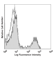
C57BL/6 mouse splenocytes were stained with CD4 (clone GK1.5) Alexa Fluor® 594 (filled histogram). The data was acquired by BD LSRFortessa™ cell analyzer equipped with Yellow-Green Laser (561 nm). -

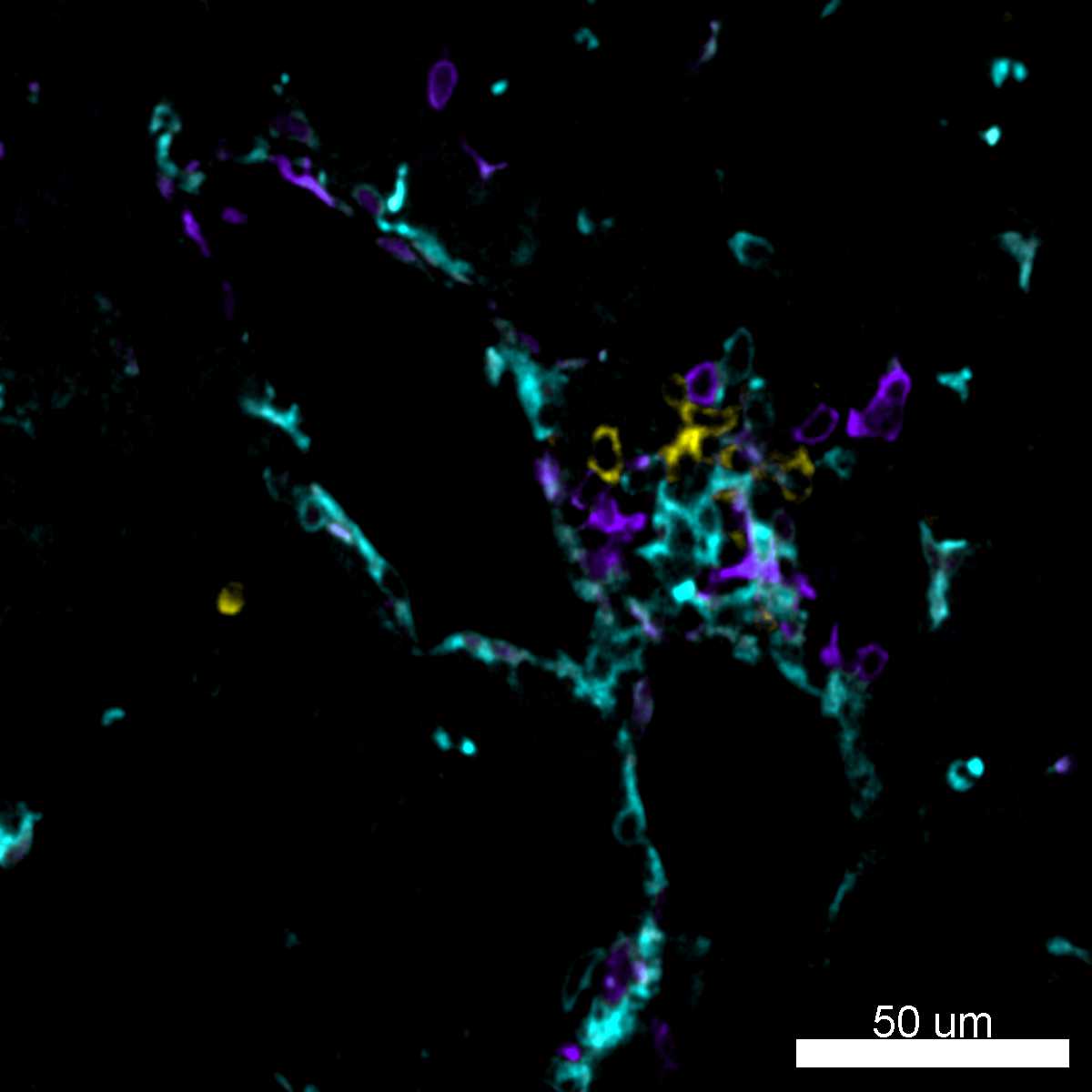
Confocal image of C57BL/6 mouse spleen sample acquired using the IBEX method of highly multiplexed antibody-based imaging: CD4 (cyan), CD8 (magenta), and IgD (blue) in Cycle 1. Tissues were prepared using ~1% (vol/vol) formaldehyde and a detergent. Following fixation, samples are immersed in 30% (wt/vol) sucrose for cryoprotection. Images are courtesy of Drs. Andrea J. Radtke and Ronald N. Germain of the Center for Advanced Tissue Imaging (CAT-I) in the National Institute of Allergy and Infectious Diseases (NIAID, NIH).
| Cat # | Size | Price | Quantity Check Availability | Save | ||
|---|---|---|---|---|---|---|
| 100446 | 100 µg | 203€ | ||||
CD4 is a 55 kD protein also known as L3T4 or T4. It is a member of the Ig superfamily, primarily expressed on most thymocytes, a subset of T cells, and weakly on macrophages and dendritic cells. It acts as a coreceptor with the TCR during T cell activation and thymic differentiation by binding MHC class II and associating with the protein tyrosin kinase, lck.
Product DetailsProduct Details
- Verified Reactivity
- Mouse
- Antibody Type
- Monoclonal
- Host Species
- Rat
- Immunogen
- Mouse CTL clone V4
- Formulation
- Phosphate-buffered solution, pH 7.2, containing 0.09% sodium azide.
- Preparation
- The antibody was purified by affinity chromatography and conjugated with Alexa Fluor® 594 under optimal conditions.
- Concentration
- 0.5 mg/ml
- Storage & Handling
- The antibody solution should be stored undiluted between 2°C and 8°C, and protected from prolonged exposure to light. Do not freeze.
- Application
-
IHC-F - Quality tested
FC - VerifiedSB - Reported in the literature, not verified in house
- Recommended Usage
-
Each lot of this antibody is quality control tested by immunohistochemistry. For immunohistochemistry on frozen tissue sections, a concentration range of 2.5-5 μg/ml is suggested. For flow cytometric staining, the suggested use of this reagent is ≤0.25 µg per million cells in 100 µl volume. It is recommended that the reagent be titrated for optimal performance for each application.
* Alexa Fluor® 594 has an excitation maximum of 590 nm, and a maximum emission of 617 nm.
Alexa Fluor® and Pacific Blue™ are trademarks of Life Technologies Corporation.
View full statement regarding label licenses - Application Notes
-
Additional reported applications (for the relevant formats) include: blocking of CD4+ T cell activation1,4,11, thymocyte costimulation3, in vitro and in vivo depletion2,5-8, blocking of egg-sperm cell adhesion1,4, immunohistochemical staining of acetone-fixed frozen sections9,10, immunoprecipitation1,2, and spatial biology (IBEX)12,13. The GK1.5 antibody is able to block CD4 mediated cell adhesion and T cell activation. Binding of GK1.5 antibody to CD4 T cells can be blocked by RM4-5 antibody, but not RM4-4 antibody. For in vivo studies or highly sensitive assays, we recommend Ultra-LEAF™ purified antibody (Cat. No. 100442) with a lower endotoxin limit than standard LEAF™ purified antibodies (Endotoxin < 0.01 EU/µg).
- Additional Product Notes
-
Iterative Bleaching Extended multi-pleXity (IBEX) is a fluorescent imaging technique capable of highly-multiplexed spatial analysis. The method relies on cyclical bleaching of panels of fluorescent antibodies in order to image and analyze many markers over multiple cycles of staining, imaging, and, bleaching. It is a community-developed open-access method developed by the Center for Advanced Tissue Imaging (CAT-I) in the National Institute of Allergy and Infectious Diseases (NIAID, NIH).
- Application References
-
- Dialynas DP, et al. 1983. J. Immunol. 131:2445. (Block, IP)
- Dialynas DP, et al. 1983. Immunol. Rev. 74:29. (IP, Deplete)
- Wu L, et al. 1991. J. Exp. Med. 174:1617. (Costim)
- Godfrey DI, et al. 1994. J. Immunol. 152:4783. (Block)
- Gavett SH, et al. 1994. Am. J. Respir. Cell. Mol. Biol. 10:587. (Deplete)
- Schuyler M, et al. 1994. Am. J. Respir. Crit. Care Med. 149:1286. (Deplete)
- Ghobrial RR, et al. 1989. Clin. Immunol. Immunopathol. 52:486. (Deplete)
- Israelski DM, et al. 1989. J. Immunol. 142:954. (Deplete)
- Zheng B, et al. 1996. J. Exp. Med. 184:1083. (IHC)
- Frei K, et al. 1997. J. Exp. Med. 185:2177. (IHC)
- Felix NJ, et al. 2007. Nat. Immunol. 8:388. (Block)
- Radtke AJ, et al. 2020. Proc Natl Acad Sci U S A. 117:33455-65. (SB) PubMed
- Radtke AJ, et al. 2022. Nat Protoc. 17:378-401. (SB) PubMed
- Product Citations
-
- RRID
-
AB_2563182 (BioLegend Cat. No. 100446)
Antigen Details
- Structure
- Ig superfamily, 55 kD
- Distribution
-
Majority of thymocytes, T cell subset
- Function
- TCR co-receptor, T cell activation
- Ligand/Receptor
- MHC class II molecule
- Cell Type
- Dendritic cells, T cells, Thymocytes, Tregs
- Biology Area
- Immunology
- Molecular Family
- CD Molecules
- Antigen References
-
1. Barclay A, et al. 1997. The Leukocyte Antigen FactsBook Academic Press.
2. Bierer BE, et al. 1989. Annu. Rev. Immunol. 7:579.
3. Janeway CA. 1992. Annu. Rev. Immunol. 10:645. - Gene ID
- 12504 View all products for this Gene ID
- UniProt
- View information about CD4 on UniProt.org
Related Pages & Pathways
Pathways


Related FAQs
- I am unable to see expression of T cell markers such as CD3 and CD4 post activation.
- TCR-CD3 complexes on the T-lymphocyte surface are rapidly downregulated upon activation with peptide-MHC complex, superantigen or cross-linking with anti-TCR or anti-CD3 antibodies. PMA/Ionomycin treatment has been shown to downregulate surface CD4 expression. Receptor downregulation is a common biological phenomenon and so make sure that your stimulation treatment is not causing it in your sample type.
- If an antibody clone has been previously successfully used in IBEX in one fluorescent format, will other antibody formats work as well?
-
It’s likely that other fluorophore conjugates to the same antibody clone will also be compatible with IBEX using the same sample fixation procedure. Ultimately a directly conjugated antibody’s utility in fluorescent imaging and IBEX may be specific to the sample and microscope being used in the experiment. Some antibody clone conjugates may perform better than others due to performance differences in non-specific binding, fluorophore brightness, and other biochemical properties unique to that conjugate.
- Will antibodies my lab is already using for fluorescent or chromogenic IHC work in IBEX?
-
Fundamentally, IBEX as a technique that works much in the same way as single antibody panels or single marker IF/IHC. If you’re already successfully using an antibody clone on a sample of interest, it is likely that clone will have utility in IBEX. It is expected some optimization and testing of different antibody fluorophore conjugates will be required to find a suitable format; however, legacy microscopy techniques like chromogenic IHC on fixed or frozen tissue is an excellent place to start looking for useful antibodies.
- Are other fluorophores compatible with IBEX?
-
Over 18 fluorescent formats have been screened for use in IBEX, however, it is likely that other fluorophores are able to be rapidly bleached in IBEX. If a fluorophore format is already suitable for your imaging platform it can be tested for compatibility in IBEX.
- The same antibody works in one tissue type but not another. What is happening?
-
Differences in tissue properties may impact both the ability of an antibody to bind its target specifically and impact the ability of a specific fluorophore conjugate to overcome the background fluorescent signal in a given tissue. Secondary stains, as well as testing multiple fluorescent conjugates of the same clone, may help to troubleshoot challenging targets or tissues. Using a reference control tissue may also give confidence in the specificity of your staining.
- How can I be sure the staining I’m seeing in my tissue is real?
-
In general, best practices for validating an antibody in traditional chromogenic or fluorescent IHC are applicable to IBEX. Please reference the Nature Methods review on antibody based multiplexed imaging for resources on validating antibodies for IBEX.
Other Formats
View All CD4 Reagents Request Custom ConjugationCustomers Also Purchased
Compare Data Across All Formats
This data display is provided for general comparisons between formats.
Your actual data may vary due to variations in samples, target cells, instruments and their settings, staining conditions, and other factors.
If you need assistance with selecting the best format contact our expert technical support team.
-
APC anti-mouse CD4

C57BL/6 mouse splenocytes were stained with CD4 (clone GK1.5... -
Biotin anti-mouse CD4

C57BL/6 mouse splenocytes were stained with biotinylated CD4... -
FITC anti-mouse CD4

C57BL/6 mouse splenocytes were stained with CD4 (clone GK1.5... -
PE anti-mouse CD4

C57BL/6 mouse splenocytes were stained with CD4 (clone GK1.5... 
C57BL/6 mouse splenocytes were stained with CD4 (clone GK1.5... -
PE/Cyanine5 anti-mouse CD4

C57BL/6 mouse splenocytes were stained with CD4 (clone GK1.5... -
Purified anti-mouse CD4

BALB/c mouse splenocytes were stained with purified CD4 (clo... -
PE/Cyanine7 anti-mouse CD4

C57BL/6 mouse splenocytes were stained with CD4 (clone GK1.5... -
APC/Cyanine7 anti-mouse CD4

C57BL/6 mouse splenocytes were stained with CD3 FITC and CD4... -
Alexa Fluor® 647 anti-mouse CD4

C57BL/6 mouse splenocytes were stained with CD4 (clone GK1.5... 
C57BL/6 mouse frozen lymph node section was fixed with 4% pa... -
Alexa Fluor® 488 anti-mouse CD4

C57BL/6 mouse splenocytes were stained with CD4 (clone GK1.5... 
C57BL/6 mouse frozen lymph node section was fixed with 4% pa... -
Pacific Blue™ anti-mouse CD4

C57BL/6 mouse splenocytes were stained with CD4 (clone GK1.5... -
Alexa Fluor® 700 anti-mouse CD4

C57BL/6 mouse splenocytes stained with CD4 (clone GK1.5) Ale... -
PerCP anti-mouse CD4

C57BL/6 mouse splenocytes were stained with CD4 (clone GK1.5... -
PerCP/Cyanine5.5 anti-mouse CD4

C57BL/6 mouse splenocytes were stained with CD3e FITC and CD... -
Brilliant Violet 421™ anti-mouse CD4

Confocal image of C57BL/6 mouse liver sample acquired using ... 
C57BL/6 mouse splenocytes were fixed with 2% paraformaldehyd... 
C57BL/6 mouse splenocytes were stained with CD3ε FITC and CD... -
Ultra-LEAF™ Purified anti-mouse CD4

C57BL/6 mouse splenocytes were stained with LEAF™ purified C... -
Alexa Fluor® 594 anti-mouse CD4

C57BL/6 mouse splenocytes were stained with CD4 (clone GK1.5... 
C57BL/6 mouse frozen lymph node section was fixed with 4% pa... 
Confocal image of C57BL/6 mouse spleen sample acquired using... -
Brilliant Violet 711™ anti-mouse CD4

C57BL/6 mouse splenocytes were stained with CD3ε FIT... 
-
Brilliant Violet 510™ anti-mouse CD4

C57BL/6 mouse splenocytes were stained with CD3ε FITC and CD... 
-
Brilliant Violet 605™ anti-mouse CD4

C57BL/6 mouse splenocytes were stained with CD3ε FIT... 
-
Brilliant Violet 785™ anti-mouse CD4

C57BL/6 mouse splenocytes were stained with CD3ε APC... 
-
PE/Dazzle™ 594 anti-mouse CD4

C57BL/6 mouse splenocytes were stained with CD3ε APC... 
-
APC/Fire™ 750 anti-mouse CD4

C57BL/6 mouse thymocytes were stained with CD8 PE and CD4 (c... 
-
GoInVivo™ Purified anti-mouse CD4
-
Brilliant Violet 750™ anti-mouse CD4

C57BL/6 mouse splenocytes were stained with CD4 (clone GK1.5... -
Brilliant Violet 650™ anti-mouse CD4

C57BL/6 mouse splenocytes were stained with CD3ε FITC and CD... -
Spark Blue™ 550 anti-mouse CD4

C57BL/6 mouse splenocytes were stained with CD4 (clone GK1.5... -
Spark NIR™ 685 anti-mouse CD4

C57BL/6 mouse splenocytes were stained with CD4 (clone GK1.5... -
KIRAVIA Blue 520™ anti-mouse CD4

C57BL/6 mouse splenocytes were stained with CD3 APC and CD4 ... -
PE/Fire™ 640 anti-mouse CD4

C57BL/6 mouse splenocytes were stained with CD3 Alexa Fluor®... -
APC/Fire™ 810 anti-mouse CD4

C57BL/6 splenocytes were stained with CD3 PE and CD4 (clone ... -
PE/Fire™ 700 anti-mouse CD4

C57BL/6 mouse splenocytes were stained with anti-mouse CD3 A... -
Spark Violet™ 538 anti-mouse CD4

C57BL/6 mouse splenocytes were stained with anti-mouse CD3ε ... -
Spark YG™ 593 anti-mouse CD4

C57BL/6 mouse splenocytes cells were stained with anti-mouse... -
Spark Blue™ 574 anti-mouse CD4 Antibody

C57BL/6 splenocytes were stained with anti-mouse CD3e APC an... -
Spark UV™ 387 anti-mouse CD4

C57BL/6 mouse splenocytes were stained with anti-mouse CD3ε ... -
Spark Blue™ 515 anti-mouse CD4

C57BL/6 mouse splenocytes were stained with anti-mouse CD3 A... -
Spark PLUS UV™ 395 anti-mouse CD4

C57BL/6 mouse splenocytes were stained with anti-mouse CD3ε ...


Follow Us