


- Clone
- BM8 (See other available formats)
- Regulatory Status
- RUO
- Other Names
- EMR1, Ly71
- Isotype
- Rat IgG2a, κ
- Ave. Rating
- Submit a Review
- Product Citations
- publications

-

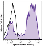
Thioglycolate-elicited Balb/c mouse peritoneal macrophages were stained with CD11b FITC and F4/80 (clone BM8) APC/Cyanine7 (left) or Rat IgG2a, κ APC/Cyanine7 isotype control (right).
| Cat # | Size | Price | Save |
|---|---|---|---|
| 123117 | 25 µg | ¥30,360 | |
| 123118 | 100 µg | ¥71,500 |
F4/80, also known as EMR1 or Ly71, is a 160 kD glycoprotein of the epidermal growth factor (EGF)-transmembrane 7 (TM7) family. F4/80 has been widely used as a murine macrophage marker. It is expressed on a majority of tissue macrophages, including macrophages in the lung, gut, peritoneal cavity, thymus, and red pulp of the spleen, Kupffer cells, Langerhans cells, microglia, and certain dendritic cells. It is not expressed on macrophages located in the T cell areas of the spleen, lymph node, or Peyer's patch. The biological ligand of F4/80 has not been identified, but it has been reported that F4/80 is required for the induction of CD8+ T cells-mediated peripheral tolerance.
Product DetailsProduct Details
- Verified Reactivity
- Mouse
- Antibody Type
- Monoclonal
- Host Species
- Rat
- Immunogen
- Murine macrophages
- Formulation
- Phosphate-buffered solution, pH 7.2, containing 0.09% sodium azide.
- Preparation
- The antibody was purified by affinity chromatography, and conjugated with APC/Cyanine7 under optimal conditions.
- Concentration
- 0.2 mg/ml
- Storage & Handling
- The antibody solution should be stored undiluted between 2°C and 8°C, and protected from prolonged exposure to light. Do not freeze.
- Application
-
FC - Quality tested
- Recommended Usage
-
Each lot of this antibody is quality control tested by immunofluorescent staining with flow cytometric analysis. For flow cytometric staining, the suggested use of this reagent is = 1.0 µg per 106 cells in 100 µl. It is recommended that the reagent be titrated for optimal performance for each application.
- Excitation Laser
-
Red Laser (633 nm)
- Application Notes
-
Additional reported applications (for the relevant formats) include: immunohistochemical staining of acetone-fixed frozen sections1,2 and formalin-fixed paraffin-embedded sections6,7, Western blotting, and spatial biology (IBEX)12,13.
- Additional Product Notes
- BioLegend is in the process of converting the name APC/Cy7 to APC/Cyanine7. The dye molecule remains the same, so you should expect the same quality and performance from our APC/Cyanine7 products. Please contact Technical Service if you have any questions.
-
Application References
(PubMed link indicates BioLegend citation) -
- Schaller E, et al. 2002. Mol. Cell. Biol. 22:8035. (IHC)
- Stevceva L, et al. 2001. BMC Clin Pathol. 1:3. (IHC)
- Kobayashi M, et al.2008. J. Leukoc. Biol. 83:1354. PubMed
- Poeckel D, et al. 2009. J. Biol Chem. 284:21077. PubMed
- Glass AM, et al. 2013. J. Immunol. 190:4830. PubMed
- Koehm S, et al. 2007. J. Allergy Clin. Immunol. 120:570. (IHC)
- Rankin AL, et al. 2010. J. Immunol. 184:1526. (IHC)
- Sasi SP, et al. 2014. J Biol Chem. 289:14178. PubMed
- Thakus VS, et al. 2014. Toxicol Lett. 230:322. PubMed
- Watson NB, et al. 2015. J Immunol. 194:2796. PubMed
- Hirakawa H, et al. 2015. PLoS One. 10:119360. PubMed
- Radtke AJ, et al. 2020. Proc Natl Acad Sci U S A. 117:33455-65. (SB) PubMed
- Radtke AJ, et al. 2022. Nat Protoc. 17:378-401. (SB) PubMed
- Product Citations
-
- RRID
-
AB_893489 (BioLegend Cat. No. 123117)
AB_893477 (BioLegend Cat. No. 123118)
Antigen Details
- Structure
- EGF-TM7 family member, 160 kD glycoprotein
- Distribution
-
Majority of tissue macrophages including peritoneal macrophages, macrophages in lung, gut, thymus and red pulp of spleen, Kupffer cells, Langerhans cells, bone marrow stromal cells, and a subset of dendritic cells
- Function
- Induction of immunological tolerance
- Cell Type
- Dendritic cells, Langerhans cells, Macrophages, Tregs
- Biology Area
- Cell Biology, Immunology, Innate Immunity, Neuroinflammation, Neuroscience
- Antigen References
-
1. Austy JM and Gordon S. 1981. Eur. J. Immunol. 11:805.
2. Hume DA, et al. 1983. J. Exp. Med. 158:1522.
3. Ruedl C, et al. 1996. Eur. J. Immunol. 26:1801.
4. McKnight AJ, et al. 1996. J. Biol. Chem. 271:486.
5. Lin HH, et al. 2005. J. Exp. Med. 201:1615. - Gene ID
- 13733 View all products for this Gene ID
- UniProt
- View information about F4/80 on UniProt.org
Related Pages & Pathways
Pathways


Related FAQs
Other Formats
View All F4/80 Reagents Request Custom ConjugationCustomers Also Purchased
Compare Data Across All Formats
This data display is provided for general comparisons between formats.
Your actual data may vary due to variations in samples, target cells, instruments and their settings, staining conditions, and other factors.
If you need assistance with selecting the best format contact our expert technical support team.
-
Brilliant Violet 605™ anti-mouse F4/80

Thioglycolate-elicited Balb/c mouse peritoneal macrophages w... -
Purified anti-mouse F4/80
-
Biotin anti-mouse F4/80

Thioglycolate-elicited Balb/c mouse peritoneal macrophages s... -
FITC anti-mouse F4/80

Thioglycolate-elicited Balb/c mouse peritoneal macrophages s... -
PE anti-mouse F4/80

Thioglycolate-elicited BALB/c mouse peritoneal macrophages s... -
PE/Cyanine5 anti-mouse F4/80
-
PE/Cyanine7 anti-mouse F4/80

Thioglycolate-elicited BALB/c mouse peritoneal macrophages s... -
APC anti-mouse F4/80

Thioglycolate-elicited BALB/c mouse peritoneal macrophages s... -
APC/Cyanine7 anti-mouse F4/80

Thioglycolate-elicited Balb/c mouse peritoneal macrophages w... -
Alexa Fluor® 488 anti-mouse F4/80

Thioglycolate-elicited Balb/c mouse peritoneal macrophages s... 
C57BL/6 mouse frozen spleen section was fixed with 4% parafo... 
Paraformaldehyde-fixed (1%), 500 µm-thick mouse spleen secti... -
Alexa Fluor® 647 anti-mouse F4/80

Thioglycolate-elicited Balb/c mouse peritoneal macrophages s... 
C57BL/6 mouse frozen spleen section was fixed with 4% parafo... 
Paraformaldehyde-fixed (1%), 500 µm-thick mouse spleen secti... -
Pacific Blue™ anti-mouse F4/80

Thioglycollate elicited Balb/c peritoneal macrophage cells s... -
PerCP anti-mouse F4/80

Thioglycolate-elicited Balb/c mouse peritoneal macrophages s... -
PerCP/Cyanine5.5 anti-mouse F4/80

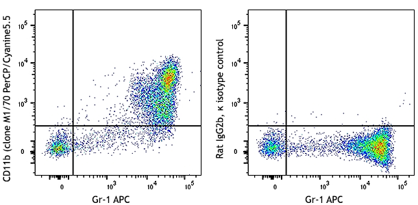
Thioglycolate-elicited Balb/c mouse peritoneal macrophages w... -
Alexa Fluor® 700 anti-mouse F4/80

Thioglycolate-elicited Balb/c peritoneal macrophages stained... -
Brilliant Violet 421™ anti-mouse F4/80

Thioglycolate-elicited Balb/c mouse peritoneal macrophages w... 
C57BL/6 mouse frozen spleen section was fixed with 4% parafo... 
Confocal image of C57BL/6 mouse spleen sample acquired using... 
Mice were injected subcutaneously with sheep red blood cells... -
Brilliant Violet 510™ anti-mouse F4/80

Thioglycolate-elicited Balb/c mouse peritoneal macrophages w... -
Alexa Fluor® 594 anti-mouse F4/80

C57BL/6 mouse frozen spleen sections were fixed with 4% para... -
Brilliant Violet 785™ anti-mouse F4/80

Thioglycolate-elicited BALB/c mouse peritoneal macrophages w... -
Purified anti-mouse F4/80 (Maxpar® Ready)

Mouse bone-marrow stained with 148Nd-anti-CD11b (M1/70) and ... -
PE/Dazzle™ 594 anti-mouse F4/80

Thioglycolate-elicited BALB/c mouse peritoneal macrophages w... -
Brilliant Violet 650™ anti-mouse F4/80

Thioglycolate-elicited Balb/c mouse peritoneal macrophages w... -
Brilliant Violet 711™ anti-mouse F4/80

Thioglycolate-elicited Balb/c mouse peritoneal macrophages w... -
APC/Fire™ 750 anti-mouse F4/80

Thioglycolate-elicited Balb/c mouse peritoneal macrophages w... -
TotalSeq™-A0114 anti-mouse F4/80
-
TotalSeq™-B0114 anti-mouse F4/80
-
TotalSeq™-C0114 anti-mouse F4/80
-
Spark YG™ 570 anti-mouse F4/80

C57BL/6 mouse frozen spleen section was fixed with 4% parafo... -
KIRAVIA Blue 520™ anti-mouse F4/80

Thioglycolate-elicited Balb/c mouse peritoneal macrophages w... -
Ultra-LEAF™ Purified anti-mouse F4/80

Thioglycollate elicited Balb/c peritoneal macrophage cells s... -
APC/Fire™ 810 anti-mouse F4/80

Thioglycolate-elicited Balb/c mouse peritoneal macrophages w... -
Spark NIR™ 685 anti-mouse F4/80

Thioglycolate-elicited BALB/c mouse peritoneal macrophages w... -
Spark Blue™ 550 anti-mouse F4/80

Thioglycolate-elicited Balb/c mouse peritoneal macrophages w... -
Brilliant Violet 570™ anti-mouse F4/80

Thioglycolate-elicited BALB/c mouse peritoneal macrophages w... -
Brilliant Violet 750™ anti-mouse F4/80

Thioglycolate-elicited BALB/c mouse peritoneal macrophages w... -
PE/Fire™ 810 anti-mouse F4/80

Thioglycolate-elicited BALB/c mouse peritoneal macrophages w...


Follow Us