 Login / Register


- Clone
- 7F11A10 (See other available formats)
- Regulatory Status
- RUO
- Other Names
- T-cell-specific transcription factor 1, transcription factor 7, TCF7
- Isotype
- Mouse IgG1, κ
- Ave. Rating
- Submit a Review
- Product Citations
- publications

-

Total cell lysate from Jurkat (lane 1), Molt-4 (lane 2), HeLa (lane 3) and EL4 (lane 4) cell lines (15 µg protein for each) were resolved by 4-12% Bis-Tris gel electrophoresis, transferred to nitrocellulose, and probed with 2 µg/mL (1:250 dilution) of purified anti-TCF1 (TCF7) antibody (clone 7F11A10) (upper blot). Proteins were visualized using chemiluminescence detection after incubation with 1:3000 dilution of HRP conjugated anti-mouse-IgG secondary antibody for the anti-TCF1 (TCF7) antibody (upper blot) 1:5000 dilution of Direct-Blot™ HRP anti-β-Actin (clone 2F1-1, lower blot). Lane M: Molecular Weight Marker -

Jurkat cells were fixed with 4% PFA for ten minutes, permeabilized with 0.5% Triton X-100 for five minutes, and blocked with 5% FBS for 30 minutes. Then the cells were stained with 2 µg/mL anti-TCF1 Antibody (clone 7F11A10) followed by Alexa Fluor® 594 (red) conjugated goat anti-mouse IgG in blocking buffer for two hours at room temperature. Actin filaments were labeled with Alexa Fluor® 488 Phalloidin (green). Nuclei were counterstained with DAPI (blue). The image was captured with a 60X objective. -

IHC staining using purified anti- TCF7 antibody (clone 7F11A10) on formalin-fixed paraffin-embedded human tonsil tissue. Following antigen retrieval using 1X Tris-EDTA (final concentration 0.05M), the tissue was incubated with 10 µg/mL of antibody overnight at 4°C, followed by incubation with 2.5 µg/mL of Alexa Fluor® 647 goat anti-mouse IgG antibody (Cat. No. 405322) for one hour at room temperature. Nuclei were counterstained with DAPI (blue) (Cat. No. 422801), and the slide was mounted with ProLong™ Gold Antifade Mountant. The images shown above (left: secondary only control; right: clone 7F11A10 at 10 µg/mL) were captured with a 40X objective. Scalebar = 50 µM.
| Cat # | Size | Price | Quantity Check Availability | Save | ||
|---|---|---|---|---|---|---|
| 655202 | 100 µg | 288 CHF | ||||
TCF1 is the first identified member of the T-cell-specific transcription factor family. It plays an important role in T cell development and differentiation. TCF1 is inactivated by association with the transcriptional repressor TLE proteins. During Wnt signaling, the transcriptional coactivator CTNNB1 accumulates and, in turn, replaces the transcriptional repressor associated with TCF1. Interaction with CTNNB1 results in transactivation of TCF1 target genes. Deletion of TCF1 causes massive apoptosis of double positive thymocyte, suggesting that TCF1 is required for thymocyte survival during T cell development. In addition to its function in thymus, TCF1 promotes T cell differentiation to Th2 cells in the periphery through transcriptional activation of GATA3.
Product DetailsProduct Details
- Verified Reactivity
- Human
- Antibody Type
- Monoclonal
- Host Species
- Mouse
- Immunogen
- Partial TCF1 recombinant protein (116-334 aa)
- Formulation
- This antibody is provided in phosphate-buffered solution, pH 7.2, containing 0.09% sodium azide.
- Preparation
- Affinity purified
- Concentration
- 0.5 mg/ml
- Storage & Handling
- Upon receipt, store undiluted between 2°C and 8°C.
- Application
-
WB - Quality tested
ICC, IHC-P - Verified - Recommended Usage
-
Each lot of this antibody is quality control tested by Western blotting. For Western blotting, the suggested use of this reagent is 0.1 - 0.5 µg per mL. For immunocytochemmistry microscopy, a concentration range of 1-5 µg/mL is recommended. For immunohistochemistry on formalin-fixed paraffin-embedded tissue sections, a concentration range of 1 - 10 µg/mL is suggested. It is recommended that the reagent be titrated for optimal performance for each application.
- Product Citations
-
- RRID
-
AB_2562103 (BioLegend Cat. No. 655202)
Antigen Details
- Structure
- 384 amino acids, predicted molecular weight of 42 kD. Contains a HMG box DNA binding domain and a CTNNB1 (β-catenin) binding domain.
- Distribution
-
Nucleus
- Function
- TCF1 is a transcription factor, involved in the canonical Wingless/Integration 1 (Wnt) signaling pathway. TCF1 is essential for survival of CD4+CD8+ double positive thymocytes and differentiation of T cells in the periphery.
- Interaction
- TCF1 interacts with CTNNB1, TLE1, TLE2, TLE3, TLE4, and AES.
- Cell Type
- Tregs
- Biology Area
- Cell Biology, Immunology, Transcription Factors
- Molecular Family
- Nuclear Markers, TCRs
- Antigen References
-
1. Mao CD, et al. 2011. Crit. Rev. Eukaryot. Gene Expr. 21:207.
2. Germar K, et al. 2011. P. Natl. Acad. Sci. USA 108:20060.
3. Wang R, et al. 2011. J. Immunol. 187:5964.
4. Weber BN, et al. 2011. Nature 476:63.
5. Jeannet G, et al. 2010. P. Natl. Acad. Sci. USA 107:9777.
6. Staal FJ, et al. 1999. Int. Immunol. 11:317. - Gene ID
- 6932 View all products for this Gene ID
- UniProt
- View information about TCF1 on UniProt.org

Related FAQs
Other Formats
View All TCF1 Reagents Request Custom Conjugation| Description | Clone | Applications |
|---|---|---|
| Purified anti-TCF1 (TCF7) | 7F11A10 | WB,ICC,IHC-P |
| Alexa Fluor® 647 anti-TCF1 (TCF7) | 7F11A10 | ICFC |
| PE anti-TCF1 (TCF7) | 7F11A10 | ICFC |
| Direct-Blot™ HRP anti-TCF1 (TCF7) | 7F11A10 | WB |
Customers Also Purchased
Compare Data Across All Formats
This data display is provided for general comparisons between formats.
Your actual data may vary due to variations in samples, target cells, instruments and their settings, staining conditions, and other factors.
If you need assistance with selecting the best format contact our expert technical support team.
-
Purified anti-TCF1 (TCF7)

Total cell lysate from Jurkat (lane 1), Molt-4 (lane 2), HeL... 
Jurkat cells were fixed with 4% PFA for ten minutes, permeab... 
IHC staining using purified anti- TCF7 antibody (clone 7F11A... -
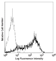
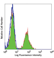
Alexa Fluor® 647 anti-TCF1 (TCF7)

Human peripheral blood lymphocytes were surface stained with... 
-
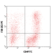
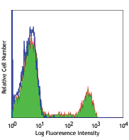
PE anti-TCF1 (TCF7)

Human peripheral blood lymphocytes were surface stained with... -
Direct-Blot™ HRP anti-TCF1 (TCF7)

Western blot analysis of 15µg cell lysates from Jurkat (high... 
Western blot analysis of 15µg cell lysates from Jurkat (high...


Follow Us