 Login / Register


- Clone
- EH12.2H7 (See other available formats)
- Regulatory Status
- RUO
- Other Names
- PD-1, PDCD1
- Isotype
- Mouse IgG1, κ
- Ave. Rating
- Submit a Review
- Product Citations
- publications

-

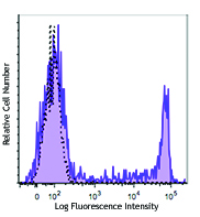
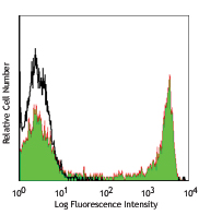
PHA-stimulated (day-3) human peripheral blood lymphocytes were stained with CD279 (clone EH12.2H7) PE (filled histogram) or mouse IgG1, κ PE (open histogram). -

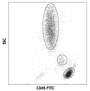
Human peripheral blood lymphocytes were stained with CD279 (clone EH12.2H7) PE and CD3 (clone UCHT1) PerCP/Cy5.5. -

Confocal image of human lymph node sample acquired using the IBEX method of highly multiplexed antibody-based imaging: IgD (blue) in Cycle 2, PD-1 (green) in Cycle 5. Tissues were prepared using ~1% (vol/vol) formaldehyde and a detergent. Following fixation, samples are immersed in 30% (wt/vol) sucrose for cryoprotection. Images are courtesy of Drs. Andrea J. Radtke and Ronald N. Germain of the Center for Advanced Tissue Imaging (CAT-I) in the National Institute of Allergy and Infectious Diseases (NIAID, NIH).
| Cat # | Size | Price | Quantity Check Availability | Save | ||
|---|---|---|---|---|---|---|
| 329905 | 25 tests | 134 CHF | ||||
| 329906 | 100 tests | 292 CHF | ||||
Programmed cell death 1 (PD-1), also known as CD279, is a 55 kD member of the immunoglobulin superfamily. CD279 contains the immunoreceptor tyrosine-based inhibitory motif (ITIM) in the cytoplasmic region and plays a key role in peripheral tolerance and autoimmune disease. CD279 is expressed predominantly on activated T cells, B cells, and myeloid cells. PD-L1 (B7-H1) and PD-L2 (B7-DC) are ligands of CD279 (PD-1) and are members of the B7 gene family. Evidence suggests overlapping functions for these two PD-1 ligands and their constitutive expression on some normal tissues and upregulation on activated antigen-presenting cells. Interaction of CD279 ligands results in inhibition of T cell proliferation and cytokine secretion.
Product DetailsProduct Details
- Verified Reactivity
- Human
- Reported Reactivity
- African Green, Baboon, Chimpanzee, Common Marmoset, Cynomolgus, Rhesus, Squirrel Monkey
- Antibody Type
- Monoclonal
- Host Species
- Mouse
- Formulation
- Phosphate-buffered solution, pH 7.2, containing 0.09% sodium azide and BSA (origin USA)
- Preparation
- The antibody was purified by affinity chromatography, and conjugated with PE under optimal conditions.
- Concentration
- Lot-specific (to obtain lot-specific concentration and expiration, please enter the lot number in our Certificate of Analysis online tool.)
- Storage & Handling
- The CD279 antibody solution should be stored undiluted between 2°C and 8°C, and protected from prolonged exposure to light. Do not freeze.
- Application
-
FC - Quality tested
SB - Reported in the literature, not verified in house - Recommended Usage
-
Each lot of this antibody is quality control tested by immunofluorescent staining with flow cytometric analysis. For flow cytometric staining, the suggested use of this reagent is 5 µl per million cells in 100 µl staining volume or 5 µl per 100 µl of whole blood.
- Excitation Laser
-
Blue Laser (488 nm)
Green Laser (532 nm)/Yellow-Green Laser (561 nm)
- Application Notes
-
Additional reported applications (for the relevant formats) include: blocking of ligand binding1-3, immunohistochemical staining of paraformaldehyde fixed frozen sections13, and spatial biology (IBEX)15,16. The LEAF™ purified antibody (Endotoxin <0.1 EU/µg, Azide-Free, 0.2 µm filtered) is recommended for functional assays (Cat. No. 329911 and 329912). For highly sensitive assays, we recommend Ultra-LEAF™ purified antibody (Cat. No. 329926) with a lower endotoxin limit than standard LEAF™ purified antibodies (Endotoxin <0.01 EU/µg).
- Additional Product Notes
-
Iterative Bleaching Extended multi-pleXity (IBEX) is a fluorescent imaging technique capable of highly-multiplexed spatial analysis. The method relies on cyclical bleaching of panels of fluorescent antibodies in order to image and analyze many markers over multiple cycles of staining, imaging, and, bleaching. It is a community-developed open-access method developed by the Center for Advanced Tissue Imaging (CAT-I) in the National Institute of Allergy and Infectious Diseases (NIAID, NIH).
-
Application References
(PubMed link indicates BioLegend citation) -
- Dorfman DM, et al. 2006 Am. J. Surg. Pathol. 30:802. (FA)
- Radziewicz H, et al. 2007. J. Virol. 81:2545. (FA)
- Velu V, et al. 2007. J. Virol. 81:5819. (FA)
- Zahn RC, et al. 2008. J. Virol. 82:11577. PubMed
- Chang WS, et al. 2008. J. Immunol. 181:6707. (FC) PubMed
- Nakamoto N, et al. 2009. PLoS Pathog. 5:e1000313. (FA)
- Jones RB, et al. 2009. J. Virol. 83:8722. (FC) PubMed
- Vojnov L, et al. 2010. J. Virol. 84:753. (FC) PubMed
- Radziewicz H, et al. 2010. J. Immunol. 184:2410. (FC) PubMed
- Monteriro P, et al. 2011. J. Immunol. 186:4618. PubMed
- Conrad J, et al. 2011. J. Immunol. 186:6871. PubMed
- Salisch NC, et al. 2010. J. Immunol. 184:476. (Rhesus reactivity)
- Li H and Pauza CD. 2015. Eur. J. Immunol. 45:298. (IHC)
- Peterson VM, et al. 2017. Nat. Biotechnol. 35:936. (PG)
- Radtke AJ, et al. 2020. Proc Natl Acad Sci USA. 117:33455-33465. (SB) PubMed
- Radtke AJ, et al. 2022. Nat Protoc. 17:378-401. (SB) PubMed
- Product Citations
-
- RRID
-
AB_940481 (BioLegend Cat. No. 329905)
AB_940483 (BioLegend Cat. No. 329906)
Antigen Details
- Structure
- Immunoglobulin superfamily
- Distribution
-
Transiently expressed on CD4- CD8- thymocytes; upregulated in thymocytes and splenic T and B lymphocytes; expressed on activated myeloid cells
- Ligand/Receptor
- B7-H1 (also known as PD-L1) and B7-DC (PD-L2)
- Cell Type
- B cells, Lymphocytes, T cells, Thymocytes, Tregs
- Biology Area
- Cancer Biomarkers, Immunology, Inhibitory Molecules
- Molecular Family
- CD Molecules, Immune Checkpoint Receptors
- Gene ID
- 5133 View all products for this Gene ID
- UniProt
- View information about CD279 on UniProt.org
Related Pages & Pathways
Pathways


Related FAQs
- What type of PE do you use in your conjugates?
- We use R-PE in our conjugates.
- If an antibody clone has been previously successfully used in IBEX in one fluorescent format, will other antibody formats work as well?
-
It’s likely that other fluorophore conjugates to the same antibody clone will also be compatible with IBEX using the same sample fixation procedure. Ultimately a directly conjugated antibody’s utility in fluorescent imaging and IBEX may be specific to the sample and microscope being used in the experiment. Some antibody clone conjugates may perform better than others due to performance differences in non-specific binding, fluorophore brightness, and other biochemical properties unique to that conjugate.
- Will antibodies my lab is already using for fluorescent or chromogenic IHC work in IBEX?
-
Fundamentally, IBEX as a technique that works much in the same way as single antibody panels or single marker IF/IHC. If you’re already successfully using an antibody clone on a sample of interest, it is likely that clone will have utility in IBEX. It is expected some optimization and testing of different antibody fluorophore conjugates will be required to find a suitable format; however, legacy microscopy techniques like chromogenic IHC on fixed or frozen tissue is an excellent place to start looking for useful antibodies.
- Are other fluorophores compatible with IBEX?
-
Over 18 fluorescent formats have been screened for use in IBEX, however, it is likely that other fluorophores are able to be rapidly bleached in IBEX. If a fluorophore format is already suitable for your imaging platform it can be tested for compatibility in IBEX.
- The same antibody works in one tissue type but not another. What is happening?
-
Differences in tissue properties may impact both the ability of an antibody to bind its target specifically and impact the ability of a specific fluorophore conjugate to overcome the background fluorescent signal in a given tissue. Secondary stains, as well as testing multiple fluorescent conjugates of the same clone, may help to troubleshoot challenging targets or tissues. Using a reference control tissue may also give confidence in the specificity of your staining.
- How can I be sure the staining I’m seeing in my tissue is real?
-
In general, best practices for validating an antibody in traditional chromogenic or fluorescent IHC are applicable to IBEX. Please reference the Nature Methods review on antibody based multiplexed imaging for resources on validating antibodies for IBEX.
Other Formats
View All CD279 Reagents Request Custom ConjugationCustomers Also Purchased
Compare Data Across All Formats
This data display is provided for general comparisons between formats.
Your actual data may vary due to variations in samples, target cells, instruments and their settings, staining conditions, and other factors.
If you need assistance with selecting the best format contact our expert technical support team.
-
Brilliant Violet 421™ anti-human CD279 (PD-1)

Human peripheral blood lymphocytes were stained with CD3 FIT... 
-
Purified anti-human CD279 (PD-1)

PHA-stimulated (day-3) human peripheral blood lymphocytes we... -
FITC anti-human CD279 (PD-1)

PHA-stimulated (day-3) human peripheral blood lymphocytes we... 
Human peripheral blood lymphocytes were stained with CD279 (... -
PE anti-human CD279 (PD-1)

PHA-stimulated (day-3) human peripheral blood lymphocytes we... 
Human peripheral blood lymphocytes were stained with CD279 (... 
Confocal image of human lymph node sample acquired using the... -
APC anti-human CD279 (PD-1)

PHA-stimulated (day-3) human peripheral blood lymphocytes we... 
Human peripheral blood lymphocytes were stained with CD279 (... -
Alexa Fluor® 647 anti-human CD279 (PD-1)

PHA-stimulated (day-3) human peripheral blood lymphocytes we... 
Human peripheral blood lymphocytes were stained with CD279 (... -
PerCP/Cyanine5.5 anti-human CD279 (PD-1)

PHA-stimulated (3-day) human peripheral blood lymphocytes we... 
Human peripheral blood lymphocytes were stained with CD3 APC... -
APC/Cyanine7 anti-human CD279 (PD-1)

PHA-stimulated (day-3) human peripheral blood lymphocytes st... -
Pacific Blue™ anti-human CD279 (PD-1)

Human peripheral blood lymphocytes were stained with CD279 (... 
PHA-stimulated (day-3) human peripheral blood lymphocytes we... -
PE/Cyanine7 anti-human CD279 (PD-1)

PHA-stimulated (day-3) human peripheral blood lymphocytes we... 
Human peripheral blood lymphocytes were stained with CD279 (... -
Purified anti-human CD279 (PD-1) (Maxpar® Ready)

Human PBMCs were incubated for 3 days in media alone (top) o... 
-
Brilliant Violet 605™ anti-human CD279 (PD-1)

Human peripheral blood lymphocytes were stained with CD3 FIT... -
Ultra-LEAF™ Purified anti-human CD279 (PD-1)
-
Brilliant Violet 711™ anti-human CD279 (PD-1)

Human peripheral blood lymphocytes were stained with CD3 FIT... -
Brilliant Violet 785™ anti-human CD279 (PD-1)

Human peripheral blood lymphocytes were stained with CD3 FIT... -
Brilliant Violet 510™ anti-human CD279 (PD-1)

PHA-stimulated (day-3) human peripheral blood lymphocytes we... -
Biotin anti-human CD279 (PD-1)

PHA-stimulated (3 days) human peripheral blood lymphocytes w... -
PE/Dazzle™ 594 anti-human CD279 (PD-1)

Human peripheral blood lymphocytes were stained with CD3 APC... 
PHA-stimulated (day 3) human peripheral blood lymphocytes st... 
-
Alexa Fluor® 488 anti-human CD279 (PD-1)

PHA-stimulated (day 3) human peripheral blood lymphocytes we... -
PerCP anti-human CD279 (PD-1)

PHA-stimulated (day 3) human peripheral blood lymphocytes we... -
GoInVivo™ Purified anti-human CD279 (PD-1)

Anti-human PD-1 inhibits the binding of PD-L1. Immobilized P... -
Brilliant Violet 650™ anti-human CD279 (PD-1)

Human peripheral blood lymphocytes were stained with CD3 FIT... 
PHA-stimulated (three days) human peripheral blood lymphocyt... 
-
Alexa Fluor® 700 anti-human CD279 (PD-1)

Human peripheral blood lymphocytes were stained with CD3 FIT... 
PHA-stimulated (three days) human peripheral blood lymphocyt... 
-
APC/Fire™ 750 anti-human CD279 (PD-1)

PHA-stimulated (day 3) human peripheral blood lymphocytes we... -
TotalSeq™-A0088 anti-human CD279 (PD-1)
-
TotalSeq™-B0088 anti-human CD279 (PD-1)
-
TotalSeq™-C0088 anti-human CD279 (PD-1)
-
Brilliant Violet 750™ anti-human CD279 (PD-1)

PHA-stimulated (day 3) human peripheral blood lymphocytes we... -
TotalSeq™-D0088 anti-human CD279 (PD-1)
-
PE/Fire™ 640 anti-human CD279 (PD-1)

PHA stimulated (day 3) human peripheral blood lymphocytes we... 
Human peripheral blood lymphocytes were stained with anti-hu... -
PE/Cyanine5 anti-human CD279 (PD-1)

PHA-stimulated (3 days) human peripheral blood lymphocytes w... -
Spark Red™ 718 anti-human CD279 (PD-1)

PHA-stimulated (3 days) human peripheral blood lymphocytes w...


Follow Us