 Login / Register


- Clone
- HP6017 (See other available formats)
- Regulatory Status
- RUO
- Other Names
- Immunoglobulin G
- Isotype
- Mouse IgG2a, κ
- Ave. Rating
- Submit a Review
- Product Citations
- publications

-

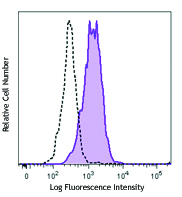
Human DLL4 transfected CHO cells were incubated with recombinant human Notch 1-IgG Fc fusion protein, and then stained with anti-human IgG Fc (clone HP6017) APC (filled histogram) or mouse IgG2a, κ APC isotype control (open histogram).
| Cat # | Size | Price | Quantity Check Availability | Save | ||
|---|---|---|---|---|---|---|
IgG Fc is a homodimer composed of the constant region of the two heavy chains that form the IgG molecule. The Fc fragment mediates opsonization, antibody dependent cellular cytotoxicity (ADCC), and complement activation through binding to Fc receptors such as CD16, CD32, CD64, and the complement factor C1.
Product DetailsProduct Details
- Verified Reactivity
- Human, Cross-Reactivity: Goat, Rabbit, Sheep, Horse
- Antibody Type
- Monoclonal
- Host Species
- Mouse
- Immunogen
- Purified human immunoglobulin
- Formulation
- Phosphate-buffered solution, pH 7.2, containing 0.09% sodium azide and 0.2% (w/v) BSA (origin USA).
- Preparation
- The antibody was purified by affinity chromatography and conjugated with APC under optimal conditions.
- Concentration
- Lot-specific (please contact technical support for concentration and total µg amount, or use our Lookup tool if you have a lot number.)
- Storage & Handling
- The antibody solution should be stored undiluted between 2°C and 8°C, and protected from prolonged exposure to light. Do not freeze.
- Application
-
FC - Quality tested
- Recommended Usage
-
Each lot of this antibody is quality control tested by immunofluorescent staining with flow cytometric analysis. For flow cytometric staining, the suggested use of this reagent is 5 µl per million cells in 100 µl staining volume or 5 µl per 100 µl of whole blood.
- Excitation Laser
-
Red Laser (633 nm)
- Application Notes
-
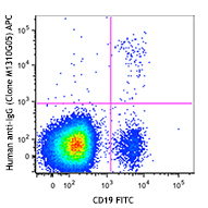
Clone HP6017 reacts with IgG (Fc fragment) of all subclasses. This antibody does not react with IgA, IgM, purified light chains, or the Fab fragment of IgG. This clone is not recommended for detection of transmembrane IgG on B cells. We recommend using clone M1310G05 as an alternative.
-
Application References
(PubMed link indicates BioLegend citation) -
- Reime CB, et al. 1984. Hybridoma 3:263.
- Jefferis R, et al. 1985. Immunol. Lett. 10:223.
- Product Citations
-
Antigen Details
- Structure
- Homodimer formed by the constant region of the IgG heavy chain
- Ligand/Receptor
- CD16, CD32, CD64
- Cell Type
- B cells, Basophils, Dendritic cells, Macrophages, Mast cells, Neutrophils, NK cells, Platelets
- Biology Area
- Adaptive Immunity, Cell Biology, Immunology, Innate Immunity
- Molecular Family
- Fc Receptors
- Antigen References
-
1. Paul WE. 2003. Fundamental Immunology. Lippincott Williams & Wilkins. Philadelphia PA.
- Gene ID
- 3500 View all products for this Gene ID
- Specificity (DOES NOT SHOW ON TDS):
- IgG Fc
- Specificity Alt (DOES NOT SHOW ON TDS):
- IgG Fc
- App Abbreviation (DOES NOT SHOW ON TDS):
- FC
- UniProt
- View information about IgG Fc on UniProt.org
Related Pages & Pathways
Pathways


Related FAQs
Customers Also Purchased
Compare Data Across All Formats
This data display is provided for general comparisons between formats.
Your actual data may vary due to variations in samples, target cells, instruments and their settings, staining conditions, and other factors.
If you need assistance with selecting the best format contact our expert technical support team.


Follow Us