 Login / Register


- Clone
- UC10-4B9 (See other available formats)
- Regulatory Status
- RUO
- Other Names
- Cytotoxic T Lymphocyte-Associated Antigen-4 (CTLA-4), Ly-56
- Isotype
- Armenian Hamster IgG
- Ave. Rating
- Submit a Review
- Product Citations
- publications

-

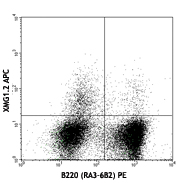
Con A+IL-2-stimulated C57BL/6 splenocytes (3 days) stained with UC10-4B9 APC and CD3e (145-2C11) PE -

Con A+IL-2-stimulated C57BL/6 splenocytes (3 days) stained with Armenian Hamster IgG isotype APC and CD3e (145-2C11) PE
| Cat # | Size | Price | Quantity Check Availability | Save | ||
|---|---|---|---|---|---|---|
| 106309 | 25 µg | 91€ | ||||
| 106310 | 100 µg | 247€ | ||||
CD152, also known as CTLA-4 or Ly-56, is a 33 kD member of the immunoglobulin superfamily. It is expressed on activated T and B lymphocytes. CD152 is similar to CD28 in amino acid sequence, structure, and genomic organization and these two receptors share common B7 family counter-receptors (B7-1, B7-2). Whereas CD28 delivers a costimulatory signal in T cell activation, CTLA-4 negatively regulates cell-mediated immune responses. CD152 is thought to play a role in the induction and maintenance of immunological tolerance as well as the development of protective immunity and thymocyte regulation.
Product DetailsProduct Details
- Verified Reactivity
- Mouse
- Antibody Type
- Monoclonal
- Host Species
- Armenian Hamster
- Immunogen
- Mouse CTLA-4-mouse IgG2a fusion protein
- Formulation
- Phosphate-buffered solution, pH 7.2, containing 0.09% sodium azide.
- Preparation
- The antibody was purified by affinity chromatography, and conjugated with APC under optimal conditions.
- Concentration
- 0.2 mg/ml
- Storage & Handling
- The antibody solution should be stored undiluted between 2°C and 8°C, and protected from prolonged exposure to light. Do not freeze.
- Application
-
FC - Quality tested
- Recommended Usage
-
Each lot of this antibody is quality control tested by immunofluorescent staining with flow cytometric analysis. For flow cytometric staining, the suggested use of this reagent is ≤1.0 µg per million cells in 100 µl volume. It is recommended that the reagent be titrated for optimal performance for each application.
- Excitation Laser
-
Red Laser (633 nm)
- Application Notes
-
The UC10-4B9 antibody can enhance T cell co-stimulation by blocking CTLA-4 interactions with the B7 co-receptors, favoring CD28 interactions. Additional reported applications (for the relevant formats) include: immunoprecipitation1, in vitro stimulation, in vitro and in vivo blocking1-4 of ligand binding, and as ELISA capture antibody5. To reduce non-specific binding to cells bearing Fc-receptors, pre-incubation of cells with anti-mouse CD16/CD32, clone 93 (Cat. No. 101301/101302), is recommended prior to immunofluorescent staining. For most successful immunofluorescent staining results, it may be important to maximize signal over background by using a relatively bright fluorochrome-antibody conjugate (Cat. No. 106306) or by using a high sensitivity, three-layer staining technique (e.g., including a biotinylated anti-Armenian hamster IgG (Cat. No. 405501) second step, followed by SAv-PE (Cat. No. 405204)). The Ultra LEAF™ purified antibody (Endotoxin < 0.01 EU/µg, Azide-Free, 0.2 µm filtered) is recommended for functional assays (Cat. No. 106327).
-
Application References
(PubMed link indicates BioLegend citation) -
- Walunas TL, et al. 1994. Immunity 1:405. (Block, IP)
- Cilio CM, et al. 1998. J. Exp. Med. 188:1239. (Block)
- Issazadeh S, et al. 1999. J. Immunol. 162:761. (Block)
- McCoy K, et al. 1997. J. Exp. Med. 186:183. (Block)
- Hsu HC, et al. 2007. J. Immunol. 178:5357. (ELISA Capture)
- Sugita S, et al. 2010. Invest. Ophthalmol. Vis. Sci. 51:5783. PubMed
- Product Citations
-
- RRID
-
AB_2230158 (BioLegend Cat. No. 106309)
AB_2087653 (BioLegend Cat. No. 106310)
Antigen Details
- Structure
- Ig superfamily, 33 kD
- Distribution
-
Activated T cells and B cells
- Function
- Negative regulator of T cell activation
- Ligand/Receptor
- CD80 (B7-1), CD86 (B7-2)
- Cell Type
- B cells, T cells, Tregs
- Biology Area
- Immunology
- Molecular Family
- CD Molecules, Immune Checkpoint Receptors
- Antigen References
-
1. Barclay A, et al. 1997. The Leukocyte Antigen FactsBook Academic Press.
2. Allison JP, et al. 1995. Science 270:932.
3. Waterhouse P, et al. 1995. Science 270:985.
4. Linsley PS, et al. 1991. J. Exp. Med. 174:561. - Gene ID
- 12477 View all products for this Gene ID
- UniProt
- View information about CD152 on UniProt.org
Related Pages & Pathways
Pathways


Related FAQs
Other Formats
View All CD152 Reagents Request Custom Conjugation| Description | Clone | Applications |
|---|---|---|
| Biotin anti-mouse CD152 | UC10-4B9 | FC |
| PE anti-mouse CD152 | UC10-4B9 | FC |
| Purified anti-mouse CD152 | UC10-4B9 | FC,IP,ELISA,Block |
| APC anti-mouse CD152 | UC10-4B9 | FC |
| Brilliant Violet 421™ anti-mouse CD152 | UC10-4B9 | FC |
| PE/Cyanine7 anti-mouse CD152 | UC10-4B9 | FC |
| PerCP/Cyanine5.5 anti-mouse CD152 | UC10-4B9 | FC |
| PE/Dazzle™ 594 anti-mouse CD152 | UC10-4B9 | FC |
| Brilliant Violet 605™ anti-mouse CD152 | UC10-4B9 | FC |
| TotalSeq™-A0388 anti-mouse CD152 | UC10-4B9 | PG |
| Ultra-LEAF™ Purified anti-mouse CD152 | UC10-4B9 | FC,IP,ELISA,Block |
| TotalSeq™-C0388 anti-mouse CD152 | UC10-4B9 | PG |
| TotalSeq™-B0388 anti-mouse CD152 | UC10-4B9 | PG |
| PE/Fire™ 640 anti-mouse CD152 | UC10-4B9 | FC |
| PE/Fire™ 810 anti-mouse CD152 | UC10-4B9 | FC |
| PE/Cyanine5 anti-mouse CD152 | UC10-4B9 | FC |
Customers Also Purchased
Compare Data Across All Formats
This data display is provided for general comparisons between formats.
Your actual data may vary due to variations in samples, target cells, instruments and their settings, staining conditions, and other factors.
If you need assistance with selecting the best format contact our expert technical support team.
-
Biotin anti-mouse CD152

Con A+IL-2-stimulated (day-2) Balb/c mouse splenocytes stain... -
PE anti-mouse CD152

Con A+IL-2-stimulated BALB/c splenocytes (day-2) were staine... -
Purified anti-mouse CD152

Con A+IL-2-stimulated C57BL/6 splenocytes (3 days) were stai... -
APC anti-mouse CD152

Con A+IL-2-stimulated C57BL/6 splenocytes (3 days) stained w... 
Con A+IL-2-stimulated C57BL/6 splenocytes (3 days) stained w... -
Brilliant Violet 421™ anti-mouse CD152

Con A+IL-2-stimulated C57BL/6 splenocytes (3 days) were stai... 
-
PE/Cyanine7 anti-mouse CD152

Con A+IL-2-stimulated C57BL/6 splenocytes (3 days) were stai... -
PerCP/Cyanine5.5 anti-mouse CD152

Con A+IL-2-stimulated C57BL/6 splenocytes (day 3) were stain... -
PE/Dazzle™ 594 anti-mouse CD152

Con A+IL-2-stimulated C57BL/6 splenocytes (3 days) were stai... 
-
Brilliant Violet 605™ anti-mouse CD152

Con A+IL-2-stimulated C57BL/6 splenocytes (three days) were ... 
-
TotalSeq™-A0388 anti-mouse CD152
-
Ultra-LEAF™ Purified anti-mouse CD152
Con A+IL-2-stimulated C57BL/6 splenocytes (3 days) were stai... -
TotalSeq™-C0388 anti-mouse CD152
-
TotalSeq™-B0388 anti-mouse CD152
-
PE/Fire™ 640 anti-mouse CD152

Con A+IL-2-stimulated C57BL/6 splenocytes (3 days) were stai... -
PE/Fire™ 810 anti-mouse CD152

Con A+IL-2-stimulated C57BL/6 splenocytes (3 days) were stai... -
PE/Cyanine5 anti-mouse CD152

Con A+IL-2 stimulated C57BL/6 splenocytes (3 days) were stai...


Follow Us