 Login / Register


- Clone
- A20004B (See other available formats)
- Regulatory Status
- RUO
- Other Names
- Cyclin Dependent Kinase 1, Cdk1, CDC28A, P34CDC2, P34 Protein Kinase
- Isotype
- Mouse IgG2b, κ
- Ave. Rating
- Submit a Review
- Product Citations
- publications

-

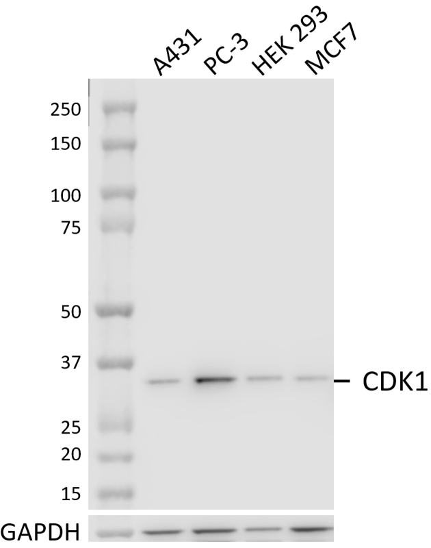
Whole cell extracts (15 µg total protein) from serum-starved HeLa cells untreated (-) or treated with 4 mM hydroxyurea for 20 hours (+) were resolved by 4-12% Bis-Tris gel electrophoresis, transferred to a PVDF membrane, treated with lambda protein phosphatase (LPP) (+) or LPP control (-) overnight at 4°C, and probed with 1.0 μg/mL (1:500 dilution) purified anti-cdc2 Phospho (Thr14) antibody (clone A20004B) for 2 hours at room temperature. Proteins were visualized by chemiluminescence detection using HRP goat anti-mouse IgG antibody (Cat. No. 405306) at a 1:3000 dilution. Western-Ready™ ECL Substrate Premium Kit (Cat. No. 426319) was used as a detection agent. Direct-Blot™ HRP anti-GAPDH antibody (Cat. No. 607904) was used as a loading control at a 1:25000 dilution (lower). Lane M: Molecular weight marker -

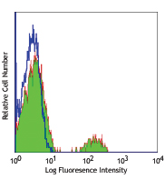
HeLa cells treated with 4 mM of hydroxyurea for 20 hours (filled histogram) (positive control) and untreated HeLa cells (open histogram) (negative control) were fixed with Fixation Buffer (Cat. No. 420801), permeabilized using True-Phos™ Perm Buffer (Cat. No. 426803), and intracellularly stained with purified anti-cdc2 Phospho (Thr14) antibody (clone A20004B) followed by PE goat anti-mouse IgG antibody (Cat. No. 405307). -

HeLa cells treated with LPP overnight at 4°C (negative) (panel A) and untreated HeLa cells (positive) (panel B) were fixed with 4% paraformaldehyde for 10 minutes, permeabilized with MeOH for 10 minutes, and blocked with 5% FBS for 60 minutes. Cells were then intracellularly stained with purified anti-cdc2 Phospho (Thr14) antibody (clone A20004B) for 2 hours at room temperature followed by incubation with Alexa Fluor® 594 goat anti-mouse IgG (Cat. No. 405326) at 2.0 µg/mL. Nuclei were counterstained with DAPI and the image was captured with a 60X objective.
| Cat # | Size | Price | Quantity Check Availability | Save | ||
|---|---|---|---|---|---|---|
| 947401 | 25 µL | 113€ | ||||
| 947402 | 100 µL | 264€ | ||||
Cyclin-dependent kinase 1 (Cdk1), also known as cell division control protein 2 (cdc2), is a key regulatory kinase essential for cell growth, proliferation, cell cycle progression, and early embryonic development. It is responsible for driving cells through G2 phase into M phase. Prior to mitosis, cdc2, along with its binding partner cyclin B1, exists in an inactive state when phosphorylated on Thr14 and Tyr15 by PKMYT1 (MYT1) and WEE1. This B1-cdc2 complex is activated during G2 and early mitosis by cdc25-mediated dephosphorylation. During mitosis, cdc2 inhibits DNA re-replication and subsequent polyploidy and cell senescence through the sequestering of cyclin A2 and suppression of the formation of CDK2-cyclin A complexes. cdc2 can function as a substitute for other cdc family members and drive the progression of the cell cycle in the event of their loss. cdc2 functions as a positive regulator of global translation, protein synthesis, and genome stability and participates in DNA damage repair, checkpoint activation, and DNA replication fork progression. It is overexpressed in several cancers including liver, breast, and colorectal cancers and has been implicated in the promotion of tumorigenesis.
Product DetailsProduct Details
- Verified Reactivity
- Human
- Antibody Type
- Monoclonal
- Host Species
- Mouse
- Immunogen
- Synthetic peptide corresponding to human cdc2 phosphorylated at threonine 14
- Formulation
- Phosphate buffer, 0.5% BSA, 0.09% NaN3
- Preparation
- The antibody was purified by affinity chromatography.
- Concentration
- 0.25 mg/mL
- Storage & Handling
- The antibody solution should be stored undiluted between 2°C and 8°C.
- Application
-
WB - Quality tested
ICC, ICFC - Verified - Recommended Usage
-
Each lot of this antibody is quality control tested by western blotting. For western blotting, the suggested dilution of this reagent is 1:4000. For immunocytochemistry, a dilution of 1:50 is recommended. For intracellular flow cytometric staining, the suggested dilution of this reagent is 1:100 per million cells in 100 µL volume. It is recommended that the reagent be titrated for optimal performance for each application.
- Application Notes
-
This clone is predicted to react with mouse cdc2 when phosphorylated at threonine 14 due to complete sequence homology between immunizing peptide and the cdc2 mouse orthologue.
This clone was tested for ICC using lambda protein phosphatase analysis and HeLa cells fixed with methanol or 4% PFA followed by permeabilization with either methanol or Triton X-100. Only PFA fixation followed by methanol permeabilization produced phospho-specific staining of cdc2. - RRID
-
AB_2904458 (BioLegend Cat. No. 947401)
AB_2904458 (BioLegend Cat. No. 947402)
Antigen Details
- Structure
- cdc2 is a 297 amino acid protein with a predicted molecular weight of 34 kD.
- Distribution
-
Ubiquitously expressed/ Mitochondria, nucleus, cytoskeleton, cytoplasm
- Function
- Serine/threonine-protein kinase/ Cell Cycle
- Interaction
- Cyclin B1, Cyclin A, PKMYT1 (MYT1)
- Biology Area
- Cell Biology, Cell Cycle/DNA Replication, Signal Transduction
- Molecular Family
- Protein Kinases/Phosphatase
- Antigen References
-
- Chow J, et al. 2011. Mol. Cell Biol. 31:1478-91.
- Diril M, et al. 2012. PNAS. 109:3826-31.
- Haneke K, et al. 2020. J. Cell. Biol. 219: e201906147.
- Liao H, et al. 2017. Oncotarget. 8:90662-73.
- Prevo R, et al. 2018. Cell Cycle. 17:1513-23.
- Gene ID
- 983 View all products for this Gene ID
- UniProt
- View information about cdc2 on UniProt.org

Related FAQs
Other Formats
View All cdc2 Reagents Request Custom Conjugation| Description | Clone | Applications |
|---|---|---|
| Purified anti-human cdc2 Phospho (Thr14) | A20004B | WB,ICC,ICFC |
| Alexa Fluor® 647 anti-human cdc2 Phospho (Thr14) | A20004B | ICFC |
| PE anti-human cdc2 Phospho (Thr14) | A20004B | ICFC |
Customers Also Purchased
Compare Data Across All Formats
This data display is provided for general comparisons between formats.
Your actual data may vary due to variations in samples, target cells, instruments and their settings, staining conditions, and other factors.
If you need assistance with selecting the best format contact our expert technical support team.
-
Purified anti-human cdc2 Phospho (Thr14)

Whole cell extracts (15 µg total protein) from serum-starved... 
HeLa cells treated with 4 mM of hydroxyurea for 20 hours (fi... 
HeLa cells treated with LPP overnight at 4°C (negative) (pan... -
Alexa Fluor® 647 anti-human cdc2 Phospho (Thr14)

HeLa cells untreated (negative control, open histogram) or t... -
PE anti-human cdc2 Phospho (Thr14)

HeLa cells treated with 4mM Hydroxyurea for 20 hours were fi... 
Hydroxyurea-treated HeLa cells (filled histogram, positive c...


Follow Us