 Login / Register


- Regulatory Status
- RUO
- Ave. Rating
- Submit a Review
- Product Citations
- publications
-

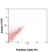
One day old splenocytes were stained with 400nM Apotracker™ Green and Annexin V Brilliant Violet 421™ in Annexin V staining buffer. -

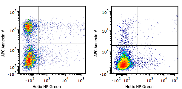
One day old splenocytes were stained with 400nM Apotracker™ Green and Helix NP™ NIR (Cat. No. 425301) in Cell Staining Buffer. -

HeLa cells were cultured for 3 days on a chamber slide coated with poly-d-lysine and then stained with Apotracker™ Green 800nM, Calcein Red-AM 0.5µM, and Helix NP™ Blue 5nM (Cat. No. 425305). Apoptotic cells (green) are Apotracker™ Green positive, live healthy cells (red) are Calcein Red-AM positive, and dead cells (blue) are Helix NP™ Blue positive.
Apotracker Green (also known as Apo-15 peptide) is a calcium-independent probe for detecting apoptotic cells. Similar to Annexin V, it detects the translocation of phosphatidylserine residues to the cell surface in a cell undergoing apoptosis. This probe can be used in conjunction with a dead cell indicator like the Helix impermeant nucleic acid stains or Zombie live/dead probes in regular cell staining buffer or PBS.
Product DetailsProduct Details
- Verified Reactivity
- Human, Mouse
- Formulation
- This product consist of dry-down Apotracker™ Green and DMSO reconstitution solution.
- Preparation
- Apotracker™ Green is prepared by dissolving the probe in methanol and drying it down.
- Storage & Handling
- Store Apotracker™ Green at -20°C
- Application
-
FC - Quality tested
Live cell imaging - Verified - Recommended Usage
-
Each lot of this product is quality control tested by immunofluorescent staining with flow cytometric analysis. For flow cytometric staining, the suggested use of this reagent is 200-800 nM per million cells in 100 µl volume. For live cell imaging, a concentration range of 600 nM-1.0 μM is recommended. It is recommended that the reagent be titrated for optimal performance for each application.
Apotracker™ Green excites at 500 nm and emits at 520 nm. The bandpass filter 530/30 is recommended for detection, although filter optimization may be required depending on other fluorophores used. Be sure to verify that your cytometer configuration and software setup are appropriate for detecting this channel. Refer to your instrument manual or manufacturer for support. The emission spectra of Apotracker™ Green is nearly identical to FITC and Alexa Fluor® 488. Therefore, we do not recommend to use them in the same panel in conventional and spectral flow cytometry. - Application Notes
-
To reconstitute the reagent to an 80 µM stock concentration, add the following volumes:
- Add 200 µL DMSO to the 200 test size
- Add 100 µL DMSO to the 100 test size
- Add 20 µL DMSO to the 20 test size
- Take the appropriate volume of reconstitute Apotracker solution to perform a 1:10 dilution with cell staining buffer.
- Add 5 µL of the diluted reagent to 1x10⁶ cells in 100 µL of an appropriate buffer, like FACS staining buffer, to achieve a 400nM staining solution. However, different cell types might require optimization of the staining solution concentration. We recommend a 200-800nM range for cells in suspension.
- Incubate at room temperature for 10-20 minutes.
- Co-stain with an appropriate dead cell stain as needed.
- Wash the cells at least 2 times prior to running on the cytometer. This reagent will be registered in the FITC channel of the flow cytometer.
- Application References
-
- Barth ND, et al. 2020. Nat Commun. 11:4027. (ICC, live cell imaging)
Antigen Details
- Biology Area
- Apoptosis/Tumor Suppressors/Cell Death, Cell Biology, Cell Proliferation and Viability
- Gene ID
- NA


Follow Us