 Login / Register


- Clone
- 53-6.7 (See other available formats)
- Regulatory Status
- RUO
- Other Names
- T8, Lyt2, Ly-2
- Isotype
- Rat IgG2a, κ
- Ave. Rating
- Submit a Review
- Product Citations
- publications

-

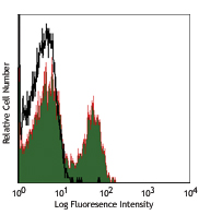
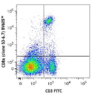
C57BL/6 mouse splenocytes were stained with CD3ε FITC and CD8a (clone 53-6.7) Brilliant Violet 421™. Quadrant gating was based on the rat IgG2a, κ Brilliant Violet 421™ isotype control. -

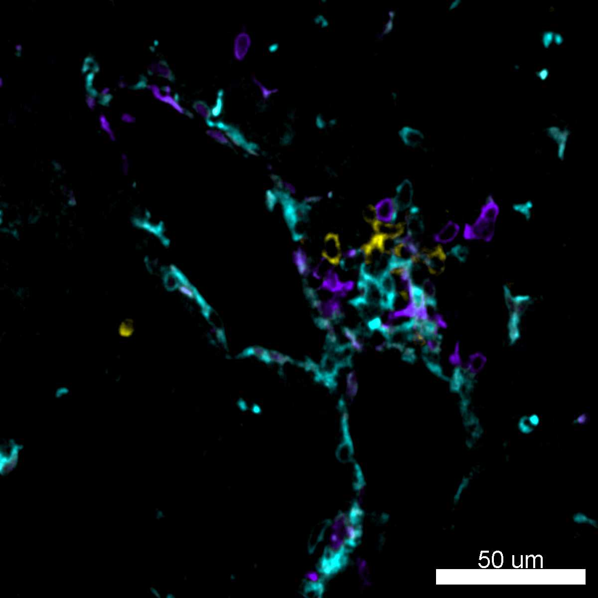
BL6 mouse lymph nodes, fixed O/N in PLP, blocked with 10% rat serum, stained with CD8a-BV421 (red), B220-Alexa Fluor® 647 (blue), and TCRβ-Alexa Fluor® 488 (green) in 1% BSA and 0.1% Tween-20 in PBS. Images were acquired with an automated widefield microscope (Nikon Eclipse Ti) and a CCD camera (QImaging Retiga 2000R). Emitted light was collected through 440/40, 525/50, and 700/75 nm bandpass filters. Images provided by Ann Haberman and Christine Podolski, Yale University. -

Confocal image of C57BL/6 mouse spleen sample acquired using the IBEX method of highly multiplexed antibody-based imaging: CD4 (cyan), CD8 (magenta), and IgD (blue) in Cycle 1. Tissues were prepared using ~1% (vol/vol) formaldehyde and a detergent. Following fixation, samples are immersed in 30% (wt/vol) sucrose for cryoprotection. Images are courtesy of Drs. Andrea J. Radtke and Ronald N. Germain of the Center for Advanced Tissue Imaging (CAT-I) in the National Institute of Allergy and Infectious Diseases (NIAID, NIH). -

Confocal image of C57BL/6 mouse thymus sample acquired using the IBEX method of highly multiplexed antibody-based imaging: CD8 (blue) in Cycle 2 and CD11c (magenta) in Cycle 3. Tissues were prepared using ~1% (vol/vol) formaldehyde and a detergent. Following fixation, samples are immersed in 30% (wt/vol) sucrose for cryoprotection. Images are courtesy of Drs. Andrea J. Radtke and Ronald N. Germain of the Center for Advanced Tissue Imaging (CAT-I) in the National Institute of Allergy and Infectious Diseases (NIAID, NIH). -

Confocal image of C57BL/6 mouse small intestine sample acquired using the IBEX method of highly multiplexed antibody-based imaging: EpCAM (magenta) in Cycle 1, CD8 (blue) in Cycle 1, and CD31 (green) in Cycle 2. Tissues were prepared using ~1% (vol/vol) formaldehyde and a detergent. Following fixation, samples are immersed in 30% (wt/vol) sucrose for cryoprotection. Images are courtesy of Drs. Andrea J. Radtke and Ronald N. Germain of the Center for Advanced Tissue Imaging (CAT-I) in the National Institute of Allergy and Infectious Diseases (NIAID, NIH). -

Confocal image of C57BL/6 mouse liver sample acquired using the IBEX method of highly multiplexed antibody-based imaging: CD8 (cyan) in Cycle 2, CD44 (blue) in Cycle 2, and NK1.1 (red) in Cycle 3. Tissues were prepared using ~1% (vol/vol) formaldehyde and a detergent. Following fixation, samples are immersed in 30% (wt/vol) sucrose for cryoprotection. Images are courtesy of Drs. Andrea J. Radtke and Ronald N. Germain of the Center for Advanced Tissue Imaging (CAT-I) in the National Institute of Allergy and Infectious Diseases (NIAID, NIH).
CD8, also known as Lyt-2, Ly-2, or T8, consists of disulfide-linked α and β chains that form the α(CD8a)/β(CD8b) heterodimer and α/α homodimer. CD8a is a 34 kD protein that belongs to the immunoglobulin family. The CD8 α/β heterodimer is expressed on the surface of most thymocytes and a subset of mature TCR α/β T cells. CD8 expression on mature T cells is non-overlapping with CD4. The CD8 α/α homodimer is expressed on a subset of γ/δ TCR-bearing T cells, NK cells, intestinal intraepithelial lymphocytes, and lymphoid dendritic cells. CD8 is an antigen co-receptor on T cells that interacts with MHC class I on antigen-presenting cells or epithelial cells. CD8 promotes T cell activation through its association with the TCR complex and protein tyrosine kinase lck.
Product DetailsProduct Details
- Verified Reactivity
- Mouse
- Antibody Type
- Monoclonal
- Host Species
- Rat
- Immunogen
- Mouse thymus or spleen
- Formulation
- Phosphate-buffered solution, pH 7.2, containing 0.09% sodium azide and BSA (origin USA).
- Preparation
- The antibody was purified by affinity chromatography and conjugated with Brilliant Violet 421™ under optimal conditions.
- Concentration
- µg sizes: 0.2 mg/mLµL sizes: lot-specific (to obtain lot-specific concentration and expiration, please enter the lot number in our Certificate of Analysis online tool.)
- Storage & Handling
- The antibody solution should be stored undiluted between 2°C and 8°C, and protected from prolonged exposure to light. Do not freeze.
- Application
-
FC - Quality tested
IHC - VerifiedSB - Reported in the literature, not verified in house
- Recommended Usage
-
Each lot of this antibody is quality control tested by immunofluorescent staining with flow cytometric analysis. For immunofluorescent staining using the µg size, the suggested use of this reagent is ≤0.5 µg per million cells in 100 µl volume. For immunofluorescent staining using µl sizes, the suggested use of this reagent is 5 µl per million cells in 100 µl staining volume or 5 µl per 100 µl of whole blood. It is recommended that the reagent be titrated for optimal performance for each application.
Brilliant Violet 421™ excites at 405 nm and emits at 421 nm. The standard bandpass filter 450/50 nm is recommended for detection. Brilliant Violet 421™ is a trademark of Sirigen Group Ltd.
Learn more about Brilliant Violet™.
This product is subject to proprietary rights of Sirigen Inc. and is made and sold under license from Sirigen Inc. The purchase of this product conveys to the buyer a non-transferable right to use the purchased product for research purposes only. This product may not be resold or incorporated in any manner into another product for resale. Any use for therapeutics or diagnostics is strictly prohibited. This product is covered by U.S. Patent(s), pending patent applications and foreign equivalents. - Excitation Laser
-
Violet Laser (405 nm)
- Application Notes
-
Clone 53-6.7 antibody competes with clone 5H10-1 antibody for binding to thymocytes3. The 53-6.7 antibody has been reported to block antigen presentation via MHC class I and inhibit T cell responses to IL-2. This antibody has also been used for depletion of CD8a+ cells. Additional reported applications (for the relevant formats) include: immunoprecipitation1,3, in vivo and in vitro cell depletion2,10,15, inhibition of CD8 T cell proliferation3, blocking of cytotoxicity3,4, immunohistochemical staining5,6 of acetone-fixed frozen sections and zinc-fixed paraffin-embedded sections, and spatial biology (IBEX)29,30. Clone 53-6.7 is not recommended for immunohistochemistry of formalin-fixed paraffin sections. The Ultra-LEAF™ purified antibody (Endotoxin < 0.01 EU/µg, Azide-Free, 0.2 µm filtered) is recommended for functional assays or in vivo studies (Cat No. 100746).
- Additional Product Notes
-
Iterative Bleaching Extended multi-pleXity (IBEX) is a fluorescent imaging technique capable of highly-multiplexed spatial analysis. The method relies on cyclical bleaching of panels of fluorescent antibodies in order to image and analyze many markers over multiple cycles of staining, imaging, and, bleaching. It is a community-developed open-access method developed by the Center for Advanced Tissue Imaging (CAT-I) in the National Institute of Allergy and Infectious Diseases (NIAID, NIH).
-
Application References
(PubMed link indicates BioLegend citation) -
- Ledbetter JA, et al. 1979. Immunol. Rev. 47:63. (IHC, IP)
- Hathcock KS. 1991. Current Protocols in Immunology. 3.4.1. (Deplete)
- Takahashi K, et al. 1992. P. Natl. Acad. Sci. USA 89:5557. (Block, IP)
- Ledbetter JA, et al. 1981. J. Exp. Med. 153:1503. (Block)
- Hata H, et al. 2004. J. Clin. Invest. 114:582. (IHC)
- Fan WY, et al. 2001. Exp. Biol. Med. 226:1045. (IHC)
- Shih FF, et al. 2006. J. Immunol. 176:3438. (FC)
- Kamimura D, et al. 2006. J. Immunol. 177:306.
- Bouwer HGA, et al. 2006. P. Natl. Acad. Sci. USA 103:5102. (FC, Deplete)
- Kao C, et al. 2005. Int. Immunol. 17:1607. PubMed
- Ko SY, et al. 2005. J. Immunol. 175:3309. (FC) PubMed
- Rasmussen JW, et al. 2006. Infect. Immun. 74:6590. PubMed
- Lee CH, et al. 2009. Clin. Cancer Res. PubMed
- Geiben-Lynn R, et al. 2008. Blood 112:4585. (Deplete) PubMed
- Kingeter LM, et al. 2008. J. Immunol. 181:6244. PubMed
- Guo Y, et al. 2008. Blood 112:480. PubMed
- Andrews DM, et al. 2008. J. Virol. 82:4931. PubMed
- Britschqui MR, et al. 2008. J. Immunol. 181:7681. PubMed
- Kenna TJ, et al. 2008. Blood 111:2091. PubMed
- Jordan JM, et al. 2008. Infect. Immun. 76:3717. PubMed
- Todd DJ, et al. 2009. J. Exp. Med. 206:2151. PubMed
- Bankoti J, et al. 2010. Toxicol. Sci. 115:422. (FC) PubMed
- Medyouf H, et al. 2010. Blood 115:1175. PubMed
- Riedl P, et al. 2009. J. Immunol. 183:370. PubMed
- Apte SH, et al. 2010. J. Immunol. 185:998. PubMed
- Bankoti J, et al. 2010. Toxicol. Sci. 115:422. (FC) PubMed
- del Rio ML, et al. 2011. Transpl. Int. 24:501. (FC) PubMed
- Cui L, et al. 2015. J Control Release. 206:220. PubMed
- Radtke AJ, et al. 2020. Proc Natl Acad Sci U S A. 117:33455-65. (SB) PubMed
- Radtke AJ, et al. 2022. Nat Protoc. 17:378-401. (SB) PubMed
- Product Citations
-
- RRID
-
AB_10897101 (BioLegend Cat. No. 100737)
AB_2562558 (BioLegend Cat. No. 100753)
AB_11204079 (BioLegend Cat. No. 100738)
Antigen Details
- Structure
- Ig superfamily, CD8α chain, 34 kD
- Distribution
-
Most thymocytes, T cell subset, some NK cells, lymphoid dendritic cells
- Function
- Co-receptor for TCR
- Ligand/Receptor
- MHC class I molecule
- Antigen References
-
1. Barclay A, et al. 1997. The Leukocyte Antigen FactsBook Academic Press.
2. Zamoyska R. 1994. Immunity 1:243.
3. Ellmeier W, et al. 1999. Annu. Rev. Immunol. 17:523. - Gene ID
- 12525 View all products for this Gene ID
- UniProt
- View information about CD8alpha on UniProt.org


Related FAQs
- What is the F/P ratio range of our BV421™ format antibody reagents?
-
It is lot-specific. On average it ranges between 2-4.
- If an antibody clone has been previously successfully used in IBEX in one fluorescent format, will other antibody formats work as well?
-
It’s likely that other fluorophore conjugates to the same antibody clone will also be compatible with IBEX using the same sample fixation procedure. Ultimately a directly conjugated antibody’s utility in fluorescent imaging and IBEX may be specific to the sample and microscope being used in the experiment. Some antibody clone conjugates may perform better than others due to performance differences in non-specific binding, fluorophore brightness, and other biochemical properties unique to that conjugate.
- Will antibodies my lab is already using for fluorescent or chromogenic IHC work in IBEX?
-
Fundamentally, IBEX as a technique that works much in the same way as single antibody panels or single marker IF/IHC. If you’re already successfully using an antibody clone on a sample of interest, it is likely that clone will have utility in IBEX. It is expected some optimization and testing of different antibody fluorophore conjugates will be required to find a suitable format; however, legacy microscopy techniques like chromogenic IHC on fixed or frozen tissue is an excellent place to start looking for useful antibodies.
- Are other fluorophores compatible with IBEX?
-
Over 18 fluorescent formats have been screened for use in IBEX, however, it is likely that other fluorophores are able to be rapidly bleached in IBEX. If a fluorophore format is already suitable for your imaging platform it can be tested for compatibility in IBEX.
- The same antibody works in one tissue type but not another. What is happening?
-
Differences in tissue properties may impact both the ability of an antibody to bind its target specifically and impact the ability of a specific fluorophore conjugate to overcome the background fluorescent signal in a given tissue. Secondary stains, as well as testing multiple fluorescent conjugates of the same clone, may help to troubleshoot challenging targets or tissues. Using a reference control tissue may also give confidence in the specificity of your staining.
- How can I be sure the staining I’m seeing in my tissue is real?
-
In general, best practices for validating an antibody in traditional chromogenic or fluorescent IHC are applicable to IBEX. Please reference the Nature Methods review on antibody based multiplexed imaging for resources on validating antibodies for IBEX.
Other Formats
View All CD8a Reagents Request Custom ConjugationCustomers Also Purchased
Compare Data Across All Formats
This data display is provided for general comparisons between formats.
Your actual data may vary due to variations in samples, target cells, instruments and their settings, staining conditions, and other factors.
If you need assistance with selecting the best format contact our expert technical support team.
-
APC anti-mouse CD8a

C57BL/6 mouse splenocytes were stained with CD8 (clone 53-6.... -
Biotin anti-mouse CD8a

C57BL/6 mouse splenocytes were stained with biotinylated CD8... -
FITC anti-mouse CD8a

C57BL/6 mouse splenocytes were stained with CD8 (clone 53-6.... -
PE anti-mouse CD8a

C57BL/6 mouse splenocytes were stained with CD8 (clone 53-6.... 
C57BL/6 mouse splenocytes were stained with CD8a (clone 53-6... -
PE/Cyanine5 anti-mouse CD8a

C57BL/6 splenocytes were stained with CD8 (clone 53-6.7) PE/... -
Purified anti-mouse CD8a

C57BL/6 mouse splenocytes were stained with purified CD8 (cl... 
C57BL/6 frozen mouse spleen section was fixed with 4% parafo... 
Fresh, frozen mouse spleen was stained with purified CD8a cl... -
PE/Cyanine7 anti-mouse CD8a

C57BL/6 mouse splenocytes were stained with anti-mouse CD3ε ... -
APC/Cyanine7 anti-mouse CD8a

C57BL/6 mouse splenocytes were stained with CD3 FITC and CD8... -
Alexa Fluor® 488 anti-mouse CD8a

C57BL/6 mouse splenocytes were stained with CD8 (clone 53-6.... 
Paraformaldehyde-fixed (1%), 500 µm-thick mouse thymus secti... -
Alexa Fluor® 647 anti-mouse CD8a

C57BL/6 mouse splenocytes were stained with CD8 (clone 53-6.... 
C57BL/6 mouse frozen lymph node section was fixed with 4% pa... 
Paraformaldehyde-fixed (1%), 500 μm-thick mouse spleen secti... 
Confocal image of C57BL/6 mouse lung sample acquired using t... -
Pacific Blue™ anti-mouse CD8a

C57BL/6 mouse splenocytes were stained with CD8 (clone 53-6.... -
Alexa Fluor® 700 anti-mouse CD8a

C57BL/6 mouse splenocytes stained with CD8 (clone 53-6.7) Al... -
PerCP/Cyanine5.5 anti-mouse CD8a

C57BL/6 mouse splenocytes were stained with CD3ε FIT... -
PerCP anti-mouse CD8a

C57BL/6 thymocytes were stained with CD8 (clone 53-6.7) PerC... -
Brilliant Violet 421™ anti-mouse CD8a

C57BL/6 mouse splenocytes were stained with CD3ε FITC and CD... 
BL6 mouse lymph nodes, fixed O/N in PLP, blocked with 10% ra... 
Confocal image of C57BL/6 mouse spleen sample acquired using... 
Confocal image of C57BL/6 mouse thymus sample acquired using... 
Confocal image of C57BL/6 mouse small intestine sample acqui... 
Confocal image of C57BL/6 mouse liver sample acquired using ... -
Brilliant Violet 570™ anti-mouse CD8a

C57BL/6 mouse splenocytes were stained with CD3 FITC and CD8... 
-
Brilliant Violet 650™ anti-mouse CD8a

C57BL/6 mouse splenocytes were stained with CD3 PE and CD8a ... -
Brilliant Violet 605™ anti-mouse CD8a

C57BL/6 mouse splenocytes were stained with CD3 FITC and CD8... -
Ultra-LEAF™ Purified anti-mouse CD8a

C57BL/6 mouse splenocytes were stained with LEAF™ purified C... -
Brilliant Violet 711™ anti-mouse CD8a

C57BL/6 mouse splenocytes were stained with CD3 PE and CD8a ... -
Brilliant Violet 785™ anti-mouse CD8a

C57BL/6 mouse splenocytes were stained with CD3 PE and CD8a ... -
Brilliant Violet 510™ anti-mouse CD8a

C57BL/6 mouse splenocytes were stained with CD3 APC and CD8a... -
Purified anti-mouse CD8a (Maxpar® Ready)

C57BL/6 mouse splenocytes stained with 147Sm-anti-CD45 (30-F... -
Alexa Fluor® 594 anti-mouse CD8a

C57BL/6 mouse frozen lymph node section was fixed with 4% pa... 
C57BL/6 mouse frozen lymph node section was fixed with 4% pa... 
Paraformaldehyde-fixed (1%), 500 µm-thick mouse thymus secti... -
PE/Dazzle™ 594 anti-mouse CD8a

C57BL/6 mouse splenocytes were stained with CD3ε FITC and C... 
-
APC/Fire™ 750 anti-mouse CD8a

C57BL/6 thymocytes were stained with CD4 PE and CD8a (clone ... 
-
GoInVivo™ Purified anti-mouse CD8a
-
TotalSeq™-A0002 anti-mouse CD8a
-
Spark Blue™ 550 anti-mouse CD8a

C57BL/6 mouse splenocytes were stained with CD8 (clone 53-6.... -
Spark NIR™ 685 anti-mouse CD8a

C57BL/6 mouse splenocytes were stained with CD8a (clone 53-6... -
TotalSeq™-C0002 anti-mouse CD8a
-
TotalSeq™-B0002 anti-mouse CD8a
-
Spark YG™ 570 anti-mouse CD8a

C57BL/6 mouse frozen spleen section was fixed with 4% parafo... -
PE/Fire™ 640 anti-mouse CD8a

C57BL/6 mouse splenocytes were stained with CD3 Alexa Fluor®... -
PE/Fire™ 700 anti-mouse CD8a

C57BL/6 mouse splenocytes were stained with anti-mouse CD3 A... -
Spark Blue™ 574 anti-mouse CD8a Antibody

C57BL/6 splenocytes were stained with anti-mouse CD3e APC an... -
Spark Violet™ 423 anti-mouse CD8a Antibody

C57BL/6 splenocytes were stained with anti-mouse CD3 PE and ... -
Spark UV™ 387 anti-mouse CD8a

C57BL/6 mouse splenocytes were stained with anti-mouse CD3ε ... -
Spark Blue™ 515 anti-mouse CD8a

C57BL/6 mouse splenocytes were stained with anti-mouse CD3ε ... -
APC/Fire™ 810 anti-mouse CD8a

C57BL/6 mouse splenocytes were stained with anti-mouse CD3&e... -
Spark Red™ 718 anti-mouse CD8a (Flexi-Fluor™)
-
PE/Fire™ 810 anti-mouse CD8a

C57BL/6 mouse splenocytes were stained with anti-mouse CD3ε ... -
Spark PLUS UV™ 395 anti-mouse CD8a

C57BL/6 mouse splenocytes were stained with anti-mouse CD3ε ...


Follow Us