 Login/Register


IBEX (Iterative Bleaching Extended multi-pleXity), is a high-content microscopy technique allowing for the analysis of dozens of parameters (more than 60) within a single tissue section. It utilizes conventional microscopes and basic laboratory techniques. While our blog gives a quick overview of the workflow and how it can be incorporated in your lab, extensive information can be found with our IBEX applications webpage below.
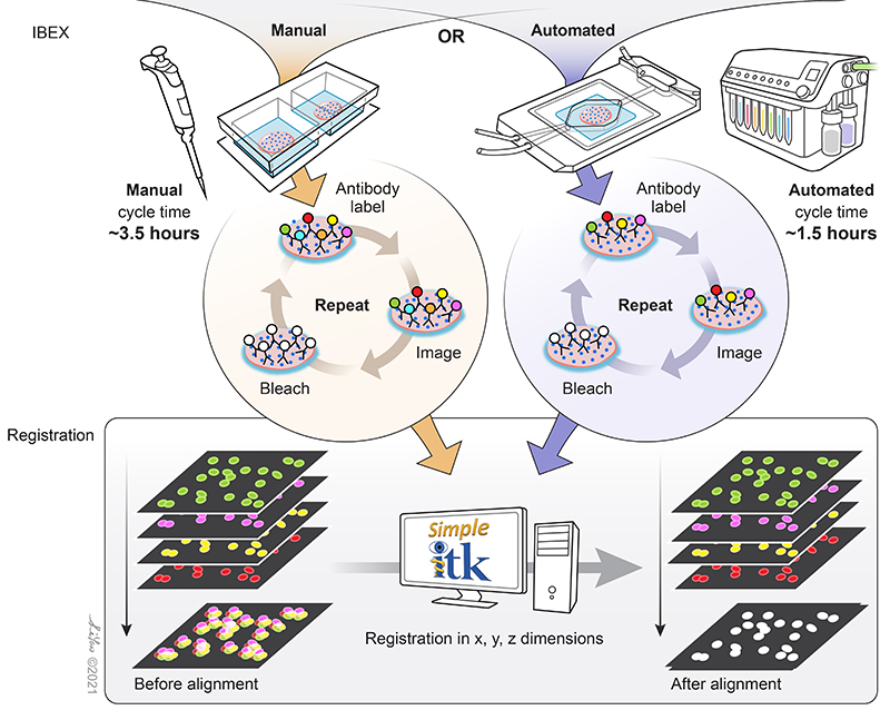
Schematic of the simple principles that allow IBEX to enable high-content imaging. Image credit: Graphical artist Li Yao
IBEX Webinar
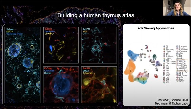
Watch a webinar with one of the developers of the technology, Andrea J. Radtke, PhD, of the Ronald Germain Lab at the Center for Advanced Tissue Imaging, NIAID/NIH. She discusses the method and how it can be used for cell atlas building and tissue phenotyping.


Follow Us