 Login / Register


- Clone
- HI30 (See other available formats)
- Regulatory Status
- RUO
- Workshop
- IV N816
- Other Names
- LCA, T200
- Isotype
- Mouse IgG1, κ
- Ave. Rating
- Submit a Review
- Product Citations
- publications

-

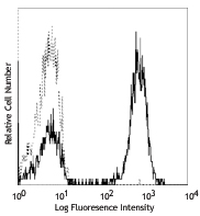
Human peripheral blood lymphocytes were stained with purified CD45 (clone HI30) (filled histogram) or purified mouse IgG1, κ isotype control (open histogram), followed by anti-mouse IgG FITC. -

Preparation and staining of formalin fixed paraffin-embedded (FFPE) human tonsil was performed. After antigen retrieval, the sample was incubated with the purified monoclonal antibody (clone HI30) at 0.5 µg/mL for 20 minutes. The Ultra Streptavidin (USA) HRP Detection Kit (Multi-Species, DAB) was used according to protocol for chromogenic detection.
| Cat # | Size | Price | Quantity Check Availability | Save | ||
|---|---|---|---|---|---|---|
| 304001 | 25 µg | 20€ | ||||
| 304002 | 100 µg | 40€ | ||||
CD45 is a 180-240 kD single chain type I membrane glycoprotein also known as leukocyte common antigen (LCA) and T200. It is a tyrosine phosphatase expressed on the plasma membrane of all hematopoietic cells, except erythrocytes and platelets. CD45 is a signaling molecule that regulates a variety of cellular processes including cell growth, differentiation, cell cycle, and oncogenic transformation. CD45 plays a critical role in T and B cell antigen receptor-mediated activation by dephosphorylating substrates including p56Lck, p59Fyn, and other Src family kinases. CD45 non-covalently associates with lymphocyte phosphatase-associated phosphoprotein (LPAP) on T and B lymphocytes. CD45 has been reported to bind galectin-1 and to be associated with several other cell surface antigens including CD1, CD2, CD3, and CD4.
Product DetailsProduct Details
- Verified Reactivity
- Human
- Reported Reactivity
- Chimpanzee
- Antibody Type
- Monoclonal
- Host Species
- Mouse
- Formulation
- Phosphate-buffered solution, pH 7.2, containing 0.09% sodium azide.
- Preparation
- The antibody was purified by affinity chromatography.
- Concentration
- 0.5 mg/ml
- Storage & Handling
- The antibody solution should be stored undiluted between 2°C and 8°C.
- Application
-
FC - Quality tested
CyTOF®, IHC-P - Verified
WB, IHC-F, ICC - Reported in the literature, not verified in house - Recommended Usage
-
Each lot of this antibody is quality control tested by immunofluorescent staining with flow cytometric analysis. For flow cytometric staining, the suggested use of this reagent is ≤ 0.5 µg per 106 cells in 100 µl volume or 100 µl of whole blood. For immunohistochemical staining on formalin-fixed paraffin-embedded tissue sections, a concentration range of 0.1 - 10 µg/ml is suggested, if the antibody is not available in a pre-diluted format. It is recommended that the reagent be titrated for optimal performance for each application.
- Application Notes
-
Additional reported applications (for the relevant formats) include: immunohistochemical staining of acetone-fixed frozen tissue sections and formalin-fixed paraffin-embedded tissue sections9, inhibition of CD45 functions4, immunofluorescence11, Western blotting3, and spatial biology (IBEX)16,17.
It was found that the HI30 clone and the 2D1 clone can cross block each other's binding. -
Application References
(PubMed link indicates BioLegend citation) -
- Knapp W, et al. 1989. Leucocyte Typing IV. Oxford University Press. New York.
- Kishihara K, et al. 1993. Cell 74:143.
- Esser M, et al. 2001. J. Virol. 75:6173. (WB)
- Yamada T, et al. 2002. J. Biol. Chem. 277:28830.
- Nagano M, et al. 2007. Blood 110:151.
- Jiang Q, et al. 2008. Blood 112:2858. PubMed
- Morozov A, et al. 2010. Clin Cancer Res. 16:5630. PubMed
- Yoshino N, et al. 2000. Exp. Anim. (Tokyo) 49:97. (FC)
- Friedman T, et al. 1999. J. Immunol. 162:5256. (IHC)
- Oeztuerk-Winder F, et al. 2012. EMBO J. 31:3431. (FC) PubMed
- Rees LE, et al. 2003. Clin. Exp. Immunol. 134:497. (IF)
- Lee J, et al. 2015. J Exp Med. 212:385. PubMed
- Breton G, et al. 2015. J Exp Med. 212:401. PubMed
- Marquardt N, et al. 2015. J Immunol. 6:2467. PubMed
- Bushway ME, et al. 2014. Biol Reprod. 90(5): 110. (IF) PubMed
- Radtke AJ, et al. 2020. Proc Natl Acad Sci USA. 117:33455-33465. (SB) PubMed
- Radtke AJ, et al. 2022. Nat Protoc. 17:378-401. (SB) PubMed
- Product Citations
-
- RRID
-
AB_314389 (BioLegend Cat. No. 304001)
AB_314390 (BioLegend Cat. No. 304002)
Antigen Details
- Structure
- Tyrosine phosphatases, type I transmembrane protein, 180-240 kD (multiple isoforms)
- Distribution
-
Hematopoietic cells, not expressed in circulating erythrocytes or platelets
- Function
- TCR and BCR mediated activation
- Ligand/Receptor
- Galectin-1, CD2, CD3, CD4
- Cell Type
- Hematopoietic stem and progenitors, Mesenchymal Stem Cells
- Biology Area
- Cell Biology, Immunology, Inhibitory Molecules, Innate Immunity, Neuroscience, Neuroscience Cell Markers, Stem Cells
- Molecular Family
- CD Molecules
- Antigen References
-
- Thomas M. 1989. Annu. Rev. Immunol. 7:339.
- Trowbridge I, et al. 1994. Annu. Rev. Immunol.12:85.
- Gene ID
- 5788 View all products for this Gene ID
- UniProt
- View information about CD45 on UniProt.org


Related FAQs
Other Formats
View All CD45 Reagents Request Custom ConjugationCustomers Also Purchased
Compare Data Across All Formats
This data display is provided for general comparisons between formats.
Your actual data may vary due to variations in samples, target cells, instruments and their settings, staining conditions, and other factors.
If you need assistance with selecting the best format contact our expert technical support team.
-
APC anti-human CD45

Human peripheral blood lymphocytes stained with HI30 APC -
Biotin anti-human CD45

Human peripheral blood lymphocytes stained with biotinylated... -
FITC anti-human CD45

Human peripheral blood lymphocytes, monocytes and granulocyt... -
PE anti-human CD45

Human peripheral blood lymphocytes stained with HI30 PE 
PE anti-human CD45 Antibody Human peripheral blood was stai... -
PE/Cyanine5 anti-human CD45

Human peripheral blood lymphocytes stained with HI30 PE/Cyan... -
Purified anti-human CD45

Human peripheral blood lymphocytes were stained with purifie... 
Preparation and staining of formalin fixed paraffin-embedded... -
APC/Cyanine7 anti-human CD45

Human peripheral blood lymphocytes stained with CD45 (clone ... -
PE/Cyanine7 anti-human CD45

Human peripheral blood lymphocytes stained with HI30 PE/Cyan... -
Alexa Fluor® 488 anti-human CD45

Human peripheral blood lymphocytes stained with HI30 Alexa F... -
Alexa Fluor® 647 anti-human CD45

Human peripheral blood lymphocytes stained with HI30 Alexa F... -
Pacific Blue™ anti-human CD45

Human peripheral blood lymphocytes stained with HI30 Pacific... -
Alexa Fluor® 700 anti-human CD45

Human peripheral blood lymphocytes stained with HI30 Alexa F... -
PerCP anti-human CD45

Human peripheral blood lymphocytes stained with HI30 PerCP -
PerCP/Cyanine5.5 anti-human CD45

Human peripheral blood lymphocytes were stained with HI30 Pe... -
Brilliant Violet 421™ anti-human CD45

Human peripheral blood lymphocytes were stained with CD45 (c... -
Brilliant Violet 570™ anti-human CD45

RBC-lysed human peripheral blood cells were stained with CD4... 
-
Brilliant Violet 510™ anti-human CD45

Human peripheral blood lymphocytes were stained with CD45 (c... -
Brilliant Violet 605™ anti-human CD45

Human peripheral blood lymphocytes were stained with CD45 (c... -
Brilliant Violet 650™ anti-human CD45

Human peripheral blood lymphocytes were stained with CD45 (c... -
Purified anti-human CD45 (Maxpar® Ready)

Human PBMCs stained with 154Sm-anti-CD45 (HI30). Data provid... -
Brilliant Violet 785™ anti-human CD45

Human peripheral blood lymphocytes were stained with CD45 (c... -
Brilliant Violet 711™ anti-human CD45

Human peripheral blood lymphocytes were stained with CD45 (c... -
PE/Dazzle™ 594 anti-human CD45

Human peripheral blood lymphocytes were stained with CD45 (c... -
Alexa Fluor® 594 anti-human CD45

Human paraffin-embedded tonsil tissue slices were prepared w... 
Confocal image of human liver sample acquired using the IBEX... -
APC/Fire™ 750 anti-human CD45

Human peripheral blood lymphocytes were stained with CD45 (c... -
Pacific Blue™ anti-human CD45

Typical results from human peripheral blood lymphocytes stai... -
APC anti-human CD45

Typical results from human peripheral blood lymphocytes stai... -
PE/Cyanine7 anti-human CD45

Typical results from human peripheral blood lymphocytes stai... -
PE/Dazzle™ 594 anti-human CD45

Typical results from human peripheral blood lymphocytes sta... -
TotalSeq™-A0391 anti-human CD45
-
TotalSeq™-B0391 anti-human CD45
-
TotalSeq™-C0391 anti-human CD45
-
PerCP/Cyanine5.5 anti-human CD45
Typical results from human peripheral blood lymphocytes stai... -
APC/Fire™ 750 anti-human CD45
Typical results from human peripheral blood lymphocytes stai... -
PE/Fire™ 640 anti-human CD45

Human peripheral blood lymphocytes were stained with CD45 (c... -
APC/Fire™ 810 anti-human CD45

Human peripheral blood lymphocytes were stained with CD45 (c... -
Spark YG™ 570 anti-human CD45

Human paraffin-embedded tonsil tissue slices were prepared w... -
PE/Fire™ 700 anti-human CD45

Human peripheral blood lymphocytes were stained with anti-hu... -
FITC anti-human CD45

Typical results from human peripheral blood lymphocytes stai... -
PerCP anti-human CD45

Typical results from human peripheral blood lymphocytes stai... -
Alexa Fluor® 660 anti-human CD45 Antibody

Human peripheral blood lymphocytes were stained with CD45 (c... -
Spark Violet™ 538 anti-human CD45

Human peripheral blood lymphocytes were stained with CD45 (c... -
Spark YG™ 593 anti-human CD45

Human peripheral blood lymphocytes were stained with CD45 (c... -
GMP APC/Fire™ 750 anti-human CD45

Typical results from human peripheral blood lymphocytes stai... -
Spark Violet™ 538 anti-human CD45

Typical results from human peripheral blood lymphocytes stai... -
GMP APC anti-human CD45

Typical results from human peripheral blood lymphocytes stai... -
Spark UV™ 387 anti-human CD45

Human peripheral blood lymphocytes were stained with anti-hu... -
GMP Pacific Blue™ anti-human CD45

Typical results from human peripheral blood lymphocytes stai... -
GMP PerCP anti-human CD45

Typical results from human peripheral blood lymphocytes stai... -
GMP FITC anti-human CD45

Typical results from human peripheral blood lymphocytes stai... -
PE anti-human CD45

Typical results from human peripheral blood lymphocytes stai... -
GMP PE/Dazzle™ 594 anti-human CD45

Typical results from human peripheral blood lymphocytes stai... -
GMP PerCP/Cyanine5.5 anti-human CD45

Typical results from human peripheral blood lymphocytes stai... -
Spark Blue™ 515 anti-human CD45

Human peripheral blood lymphocytes were stained with anti-hu... -
TotalSeq™-D0391 anti-human CD45
-
GMP PE/Cyanine7 anti-human CD45

Typical results from human peripheral blood lymphocytes stai... -
Spark Violet™ 500 anti-human CD45

Human peripheral blood lymphocytes were stained with anti-hu... -
GMP PE anti-human CD45

Typical results from human peripheral blood lymphocytes stai... -
PE/Fire™ 810 anti-human CD45

Human peripheral blood lymphocytes were stained with anti-hu... -
Spark PLUS UV™ 395 anti-human CD45

Human peripheral blood lymphocytes were stained with anti-hu... 
Human peripheral blood cells were stained with anti-human CD... -
Spark NIR™ 685 anti-human CD45

Human peripheral blood leukocytes were stained with anti-hum... -
Spark Violet™ 423 anti-human CD45

Human peripheral blood leukocytes were stained with anti-hum...


Follow Us