 Login / Register


- Clone
- 7C2C34 (See other available formats)
- Regulatory Status
- RUO
- Other Names
- Spleen focus forming virus (SFFV) proviral integration oncogene, Sfpi1, Transcription factor PU.1, Dis1, PU.1, Dis-1, Sfpi1, Spi-1, Sfpi-1, Tcfpu1, Tfpu.1
- Isotype
- Rat IgG2a, κ
- Ave. Rating
- Submit a Review
- Product Citations
- publications

-

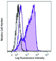
C57BL/6 splenocytes were surface stained with CD19 APC and then treated with True-Nuclear™ Transcription Factor Buffer Set. Cells were intracellularly stained with SPI1 (clone 7C2C34) PE (top) or rat IgG2a, κ PE isotype control (bottom). -

| Cat # | Size | Price | Quantity Check Availability | Save | ||
|---|---|---|---|---|---|---|
| 681307 | 25 µg | 117€ | ||||
| 681308 | 100 µg | 259€ | ||||
SPI1 is a transcription factor that belongs to the E26-transformation-specific (ETS) family and is exclusively expressed in hematopoietic cells. SPI1 regulates cell fate decisions during the differentiation of hematopoietic stem cells, which is crucial for the development of lymphoid and myeloid cell lineages. SPI1-deficient mice lack macrophages, neutrophils, and B lymphocytes, and they die before or shortly after birth. Abnormally regulated expression of SPI1 can lead to developmental defects as well as malignancy. The overexpression of SPI1 blocks erythroid differentiation and inhibits cell death. Mice carrying a mutant SPI1 allele shows decreased SPI1 expression and develops acute myeloid leukaemia (AML), which suggests that SPI1 plays a role in oncogenesis.
Product DetailsProduct Details
- Verified Reactivity
- Mouse
- Antibody Type
- Monoclonal
- Host Species
- Rat
- Immunogen
- Full length SPI1 recombinant protein expressed in E. coli.
- Formulation
- Phosphate-buffered solution, pH 7.2, containing 0.09% sodium azide.
- Preparation
- The antibody was purified by affinity chromatography and conjugated with PE under optimal conditions.
- Concentration
- 0.2 mg/ml
- Storage & Handling
- The antibody solution should be stored undiluted between 2°C and 8°C, and protected from prolonged exposure to light. Do not freeze.
- Application
-
ICFC - Quality tested
- Recommended Usage
-
Each lot of this antibody is quality control tested by intracellular flow cytometry using our True-Nuclear™ Transcription Factor Staining Protocol. For flow cytometric staining, the suggested use of this reagent is ≤ 0.125 µg per million cells in 100 µL volume. It is recommended that the reagent be titrated for optimal performance for each application.
- Excitation Laser
-
Blue Laser (488 nm)
Green Laser (532 nm)/Yellow-Green Laser (561 nm)
- Application Notes
-
NOTE: For flow cytometric staining with this clone, True-Nuclear™ Transcription Factor Buffer Set (Cat. No. 424401) offers improved staining and is highly recommended.
- Product Citations
-
- RRID
-
AB_2629617 (BioLegend Cat. No. 681307)
AB_2629618 (BioLegend Cat. No. 681308)
Antigen Details
- Structure
- 272 amino acids with a predicted molecular weight of 31.3 kD. It contains a C-terminal ETS domain that is responsible for DNA binding.
- Distribution
-
Nucleus.
- Function
- SPI1 is a transcription factor that is required for the development of lymphoid and myeloid cells.
- Interaction
- SPI1 interacts with RUNX1, CEBPD, NONO, SPIB, and GFI1.
- Biology Area
- Cell Biology, Immunology, Transcription Factors
- Molecular Family
- Nuclear Markers
- Antigen References
-
1. Hikami K, et al. 2011. Arthritis Rheum. 63:755.
2. Zakrzewska A, et al. 2010. Blood 116:e1.
3. Pham TH, et al. 2013. Nucleic Acids Res. 41:6391.
4. Pospisil V, et al. 2011. EMBO J. 30:4450.
5. Zarnegar MA, et al. 2010. Mol. Cell Biol. 30:4922.
6. Rimmele P, et al. 2010. Cancer Res. 70:6757. - Gene ID
- 20375 View all products for this Gene ID
- UniProt
- View information about SPI1 on UniProt.org


Related FAQs
- What type of PE do you use in your conjugates?
- We use R-PE in our conjugates.
Other Formats
View All SPI1 Reagents Request Custom Conjugation| Description | Clone | Applications |
|---|---|---|
| Purified anti-SPI1 (PU.1) | 7C2C34 | WB,ICC |
| Alexa Fluor® 647 anti-SPI1 (PU.1) | 7C2C34 | ICFC |
| Alexa Fluor® 488 anti-SPI1 (PU.1) | 7C2C34 | ICFC |
| PE anti-SPI1 (PU.1) | 7C2C34 | ICFC |
Customers Also Purchased
Compare Data Across All Formats
This data display is provided for general comparisons between formats.
Your actual data may vary due to variations in samples, target cells, instruments and their settings, staining conditions, and other factors.
If you need assistance with selecting the best format contact our expert technical support team.
-
Purified anti-SPI1 (PU.1)

Total cell lysates (15 µg protein) from THP1 (lane 1) and Ra... 
Raw 264.7 cells were stained with purified anti-SPI1 (clone ... -
Alexa Fluor® 647 anti-SPI1 (PU.1)

C57BL/6 splenocytes were surface stained with CD19 PE and th... 
-
Alexa Fluor® 488 anti-SPI1 (PU.1)

C57BL/6 splenocytes were surface stained with CD19 APC and t... 
-
PE anti-SPI1 (PU.1)

C57BL/6 splenocytes were surface stained with CD19 APC and t... 


Follow Us