 Login / Register


- Clone
- 67A4 (See other available formats)
- Regulatory Status
- RUO
- Other Names
- E-Cadherin, cadherin-1, and UVO
- Isotype
- Mouse IgG1, κ
- Ave. Rating
- Submit a Review
- Product Citations
- publications

-

Human paraffin-embedded colon cancer tissue slices were prepared with a standard protocol of deparaffinization and rehydration. Antigen retrieval was done with Tris-Buffered Saline 1X (1.0M, pH 7.4) at 95°C for 40 minutes. Tissue was washed with PBS/0.05% Tween 20 twice for five minutes and blocked with 5% FBS and 0.2% gelatin for 30 minutes. Then, the tissue was stained with 10 µg/mL of anti-human CD324 (clone 67A4) Alexa Fluor® 594 (red) at 4°C overnight. Nuclei were counterstained with DAPI (blue). The image was captured with a 10X objective. -

Human paraffin-embedded skin tissue slices were prepared with a standard protocol of deparaffinization and rehydration. Antigen retrieval was done with Tris-Buffered Saline 1X (1.0M, pH 7.4) at 95°C for 40 minutes. Tissue was washed with PBS/0.05% Tween 20 twice for five minutes and blocked with 5% FBS and 0.2% gelatin for 30 minutes. Then, the tissue was stained with 10 µg/mL of anti-human CD324 (clone 67A4) Alexa Fluor® 594 (red) at 4°C overnight. Nuclei were counterstained with DAPI (blue). The image was captured with a 10X objective. -

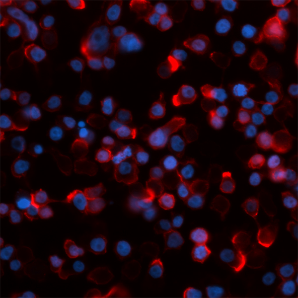
Human colorectal adenocarcinoma cell line HT-29 was fixed with 1% paraformaldahyde (PFA) and blocked with 5% FBS for 30 minutes. Then the cells were stained with 10 µg/ml anti-human CD324 (clone 67A4) Alexa Fluor® 594 (red). Nuclei were counterstained with DAPI (blue). The image was acquired with a 40X objective.
| Cat # | Size | Price | Quantity Check Availability | Save | ||
|---|---|---|---|---|---|---|
| 324118 | 100 µg | 165€ | ||||
The 67A4 antibody recognizes human CD324 also known as E-cadherin, cadherin-1, and UVO. CD324, a member of the cadherin superfamily, is a calcium-dependent, transmembrane cell-cell adhesion glycoprotein composed of 4 extracellular cadherin repeats and a highly conserved cytoplasmic tail region with a predicted molecular weight of approximately 100 kD. CD324 is widely expressed in epithelial cells in the colon, uterus, liver, keratinocytes, brain, heart, muscle, kidney, and pancreas, as well as erythroid cells. CD324 functions as a cell adhesion molecule involved in development, bacterial pathogenesis, and tumor invasion. In bacterial pathogenesis, the ectodomain of CD324 mediates bacterial adhesion to mammalian cells, while the cytoplasmic domain is required for internalization. CD324 binds to the αEβ7 integrin to mediate cell adhesion and also interacts with a number of intracellular proteins including including erbin, ezrin, caspase-3, caspase 8, β-catenin, presenilin 1, casein kinase II , as well as other extracellular proteins including the EGF receptor. CD324 is phosphorylated on multiple residues (S857, S866, S870, S872), and can be proteolytically cleaved at reside D769 by caspase-3. The 67A4 antibody has been shown to be useful for flow cytometry.
Product DetailsProduct Details
- Verified Reactivity
- Human
- Antibody Type
- Monoclonal
- Host Species
- Mouse
- Immunogen
- T-47D cells
- Formulation
- Phosphate-buffered solution, pH 7.2, containing 0.09% sodium azide.
- Preparation
- The antibody was purified by affinity chromatography and conjugated with Alexa Fluor® 594 under optimal conditions.
- Concentration
- 0.5 mg/ml
- Storage & Handling
- The antibody solution should be stored undiluted between 2°C and 8°C, and protected from prolonged exposure to light. Do not freeze.
- Application
-
IHC-P - Quality tested
ICC - Verified - Recommended Usage
-
Each lot of this antibody is quality control tested by formalin-fixed paraffin-embedded immunohistochemical staining. For immunohistochemistry, a concentration range of 2.5 - 5.0 µg/ml is suggested. For immunocytochemistry, a concentration range of 2.5 - 10 μg/ml is recommended. It is recommended that the reagent be titrated for optimal performance for each application.
It is also recommended using EDTA-based solutions for dissociating attachment-dependent cell lines.
* Alexa Fluor® 594 has an excitation maximum of 590 nm, and a maximum emission of 617 nm.
Alexa Fluor® and Pacific Blue™ are trademarks of Life Technologies Corporation.
View full statement regarding label licenses - Application References
-
- Armeanu S, et al. 1995. J. Cell Biol. 131:243.
- Bühring HJ, et al. 1996. Leukemia 10:106.
- Yauch RL, et al. 2005. Clin. Cancer Res. 11:8686. (WB)
- Oeztuerk-Winder F, et al. 2012. EMBO J. 31:3431. (FC) PubMed
- Ardehali R, et al. 2013. PNAS. 110:3405. PubMed
- Rasanen K, et al. 2013. Mol Cell Protecomics. 12:3778. PubMed
- Chaudhury A, et al. 2014. Nucleic Acids Res. 42:86. PubMed
- Milne P, et al. 2015. Blood. 125:470. PubMed
- RRID
-
AB_2563211 (BioLegend Cat. No. 324118)
Antigen Details
- Structure
- Member of the cadherin superfamily. Calcium-dependent, transmembrane cell-cell adhesion glycoprotein composed of 4 extracellular cadherin repeats and a highly conserved cytoplasmic tail region. Predicted molecular weight approximately 100 kD.
- Distribution
-
Widely expressed in epithelial cells in the colon, uterus, liver, keratinocytes, brain, heart, muscle, kidney, and pancreas, as well as erythroid cells
- Function
- Cell adhesion molecule involved in development, bacterial pathogenesis, and tumor invasion. The ectodomain of CD324 mediates bacterial adhesion to mammalian cell, while the cytoplasmic domain is required for internalization.
- Interaction
- Interacts with a variety of proteins including erbin, ezrin, caspase-3, caspase 8, EGF receptor, β-catenin, presenilin 1, casein kinase II, and others
- Ligand/Receptor
- αEβ7 integrin
- Modification
- Phosphorylated on multiple residues (S857, S866, S870, S872), proteolytically cleaved at reside D769 by caspase-3.
- Cell Type
- Embryonic Stem Cells, Epithelial cells, Erythrocytes
- Biology Area
- Cell Biology, Immunology, Neuroscience, Stem Cells, Synaptic Biology
- Molecular Family
- Adhesion Molecules, CD Molecules
- Antigen References
-
1. Overduin M, et al. 1995. Science 267:386.
2. Boggon TJ, et al. 2002. Science 296:1303.
3. Berx G, et al. 1995. EMBO J. 14:6107.
4. Perl AK, et al. 1998. Nature 392:190. - Gene ID
- 999 View all products for this Gene ID
- UniProt
- View information about CD324 on UniProt.org
Related Pages & Pathways
Pathways


Related FAQs
Other Formats
View All CD324 Reagents Request Custom Conjugation| Description | Clone | Applications |
|---|---|---|
| Purified anti-human CD324 (E-Cadherin) | 67A4 | FC,ICC,WB |
| FITC anti-human CD324 (E-Cadherin) | 67A4 | FC |
| PE anti-human CD324 (E-Cadherin) | 67A4 | FC |
| APC anti-human CD324 (E-Cadherin) | 67A4 | FC |
| Alexa Fluor® 488 anti-human CD324 (E-Cadherin) | 67A4 | FC |
| Alexa Fluor® 647 anti-human CD324 (E-Cadherin) | 67A4 | FC |
| PerCP/Cyanine5.5 anti-human CD324 (E-Cadherin) | 67A4 | FC |
| PE/Cyanine7 anti-human CD324 (E-Cadherin) | 67A4 | FC |
| Alexa Fluor® 594 anti-human CD324 (E-Cadherin) | 67A4 | IHC-P,ICC |
| APC/Fire™ 750 anti-human CD324 (E-Cadherin) | 67A4 | FC |
| Biotin anti-human CD324 (E-Cadherin) | 67A4 | FC |
| TotalSeq™-A0135 anti-human CD324 (E-Cadherin) | 67A4 | PG |
| TotalSeq™-C0135 anti-human CD324 (E-Cadherin) | 67A4 | PG |
| TotalSeq™-B0135 anti-human CD324 (E-Cadherin) | 67A4 | PG |
Customers Also Purchased
Compare Data Across All Formats
This data display is provided for general comparisons between formats.
Your actual data may vary due to variations in samples, target cells, instruments and their settings, staining conditions, and other factors.
If you need assistance with selecting the best format contact our expert technical support team.
-
Purified anti-human CD324 (E-Cadherin)

Human colon carcinoma cell line (HT29) stained with purified... 
BT474 breast cancer cells were stained with anti-human CD324... 
Total cell lysate (15 µg) from HeLa (negative control, lane... -
FITC anti-human CD324 (E-Cadherin)

Human colon carcinoma cell line (HT29) stained with CD324 (c... -
PE anti-human CD324 (E-Cadherin)

Human colon carcinoma cell line (HT29) stained with CD324 (c... -
APC anti-human CD324 (E-Cadherin)

Human colon carcinoma cell line (HT29) stained with CD324 (c... -
Alexa Fluor® 488 anti-human CD324 (E-Cadherin)

Human colon carcinoma cell line (HT29) stained with CD324 (c... -
Alexa Fluor® 647 anti-human CD324 (E-Cadherin)

Human colon carcinoma cell line (HT29) stained with CD324 (c... -
PerCP/Cyanine5.5 anti-human CD324 (E-Cadherin)

Human colon carcinoma cell line HT-29 was stained with CD324... -
PE/Cyanine7 anti-human CD324 (E-Cadherin)

Human colon carcinoma cell line (HT29) was stained with CD32... -
Alexa Fluor® 594 anti-human CD324 (E-Cadherin)

Human paraffin-embedded colon cancer tissue slices were prep... 
Human paraffin-embedded skin tissue slices were prepared wit... 
Human colorectal adenocarcinoma cell line HT-29 was fixed wi... -
APC/Fire™ 750 anti-human CD324 (E-Cadherin)

Human colon carcinoma cell line HT-29 was stained with CD324... -
Biotin anti-human CD324 (E-Cadherin)

Human colon carcinoma cell line HT-29 was stained with bioti... -
TotalSeq™-A0135 anti-human CD324 (E-Cadherin)
-
TotalSeq™-C0135 anti-human CD324 (E-Cadherin)
-
TotalSeq™-B0135 anti-human CD324 (E-Cadherin)


Follow Us