 Login / Register


- Clone
- W19063E (See other available formats)
- Regulatory Status
- RUO
- Other Names
- Tumor necrosis factor-α, Cachectin, Necrosin, Macrophage cytotoxic factor (MCF), Differentiation inducing factor (DIF), TNFSF2
- Isotype
- Rat IgG1, κ
- Ave. Rating
- Submit a Review
- Product Citations
- publications
-
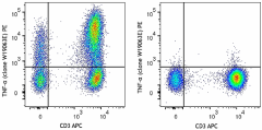
Human peripheral blood lymphocytes were left un-stimulated (right) or stimulated with PMA and Ionomycin for 6 hours in the presence of monensin (left). Cells were stained with anti-human CD3 APC. After fixation and permeabilization, cells were intracellularly stained with TNF-α (clone W19063E) PE.
| Cat # | Size | Price | Quantity Check Availability | Save | ||
|---|---|---|---|---|---|---|
| 376203 | 25 tests | 79€ | ||||
| 376204 | 100 tests | 195€ | ||||
TNF-α is secreted by macrophages, monocytes, neutrophils, T cells (principally CD4+), and NK cells. Many transformed cell lines also secrete TNF-α. Monomeric human TNF-α is a 157 amino acid protein (non-glycosylated) with a reported molecular weight of 17 kD. TNF-α forms multimeric complexes; stable trimers are most common in solution. A 26 kD membrane form of TNF-α has also been described. TNF-α binding to surface receptors elicits a wide array of biological activities including: cytolysis and cytostasis of many tumor cell lines in vitro, hemorraghic necrosis of tumors in vivo, increased fibroblast proliferation, and enhanced chemotaxis and phagocytosis in neutrophils.
Product DetailsProduct Details
- Verified Reactivity
- Human
- Antibody Type
- Monoclonal
- Host Species
- Rat
- Formulation
- Phosphate-buffered solution, pH 7.2, containing 0.09% sodium azide and BSA (origin USA)
- Preparation
- The antibody was purified by affinity chromatography and conjugated with PE under optimal conditions.
- Concentration
- Lot-specific (to obtain lot-specific concentration and expiration, please enter the lot number in our Certificate of Analysis online tool.)
- Storage & Handling
- The antibody solution should be stored undiluted between 2°C and 8°C, and protected from prolonged exposure to light. Do not freeze.
- Application
-
ICFC - Quality tested
- Recommended Usage
-
Each lot of this antibody is quality control tested by intracellular immunofluorescent staining with flow cytometric analysis. For flow cytometric staining, the suggested use of this reagent is 5 µL per million cells in 100 µL staining volume or 5 µL per 100 µL of whole blood. It is recommended that the reagent be titrated for optimal performance for each application.
- Excitation Laser
-
Blue Laser (488 nm)
Green Laser (532 nm)/Yellow-Green Laser (561 nm)
- RRID
-
AB_2894502 (BioLegend Cat. No. 376203)
AB_2894502 (BioLegend Cat. No. 376204)
Antigen Details
- Structure
- TNF superfamily; dimer/trimer; 17 kD (Mammalian)
- Distribution
-
Activated monocytes, neutrophils, macrophages, T cells, B cells, NK cells, LAK cells
- Function
- Type II integral membrane protein processed by TACE for secretion; upregulated by interferons, IL-2, GM-CSF, substance P, bradykinin, PAF, immune complexes, cyclooxygenase; downregulated by IL-6, TGF-β, vitamin D3, prostaglandin E2, PAF antagonists
- Interaction
- Monocytes, neutrophils, macrophages, T cells, fibroblasts, endothelial cells, osteoclasts, adipocytes, astroglia, microglia
- Ligand/Receptor
- TNFRSF1A (TNF-R1, CD120a, TNFR-p60 Type β, p55); TNFRSF1B (TNF-R2, CD120b, TNFR-p80 Type A, p75)
- Cell Type
- B cells, Granulocytes, Lymphocytes, Macrophages, Monocytes, NK cells, NKT cells, T cells, Th1, Th17
- Biology Area
- Adaptive Immunity, Cell Death, Immunology, Innate Immunity
- Molecular Family
- Cytokines/Chemokines
- Antigen References
-
- Volova L.T, et al. 2020. Biomolecules. 10:1563.
- Szondy Z, et al. 2017. Pharmacological Research. 115:124-132.
- Gene ID
- 7124 View all products for this Gene ID
- UniProt
- View information about TNF-alpha on UniProt.org


Related FAQs
- What type of PE do you use in your conjugates?
- We use R-PE in our conjugates.
Other Formats
View All TNF-α Reagents Request Custom Conjugation| Description | Clone | Applications |
|---|---|---|
| PE anti-human TNF-α | W19063E | ICFC |
| APC anti-human TNF-α | W19063E | ICFC |
| FITC anti-human TNF-α | W19063E | ICFC |
| PE/Cyanine7 anti-human TNF-α | W19063E | ICFC |
| Purified anti-human TNF-α | W19063E | ICFC |
| Brilliant Violet 650™ anti-human TNF-α | W19063E | ICFC |
| Brilliant Violet 421™ anti-human TNF-α | W19063E | ICFC |
| PerCP/Fire™ 806 anti-human TNF-α | W19063E | ICFC |
| Spark Blue™ 515 anti-human TNF-α | W19063E | ICFC |
| Spark UV™ 387 anti-human TNF-α | W19063E | ICFC |
Compare Data Across All Formats
This data display is provided for general comparisons between formats.
Your actual data may vary due to variations in samples, target cells, instruments and their settings, staining conditions, and other factors.
If you need assistance with selecting the best format contact our expert technical support team.
-
PE anti-human TNF-α
Human peripheral blood lymphocytes were left un-stimulated (... -
APC anti-human TNF-α

Human peripheral blood lymphocytes were stimulated with Cell... -
FITC anti-human TNF-α

Human peripheral blood lymphocytes were stimulated with Cell... -
PE/Cyanine7 anti-human TNF-α

Human peripheral blood lymphocytes were stimulated with Cell... -
Purified anti-human TNF-α

Human peripheral blood lymphocytes were left un-stimulated (... -
Brilliant Violet 650™ anti-human TNF-α

Human peripheral blood lymphocytes were stimulated with Cell... -
Brilliant Violet 421™ anti-human TNF-α

Human peripheral blood lymphocytes were left un-stimulated (... -
PerCP/Fire™ 806 anti-human TNF-α

Human peripheral blood lymphocytes were left un-stimulated (... -
Spark Blue™ 515 anti-human TNF-α

Human peripheral blood lymphocytes were left un-stimulated (... -
Spark UV™ 387 anti-human TNF-α

Human peripheral blood lymphocytes were left un-stimulated (...


Follow Us