 Login / Register


- Regulatory Status
- RUO
- Other Names
- Live/Dead Staining, Dead cell exclusion dye, Cell viability dye
- Ave. Rating
- Submit a Review
- Product Citations
- publications

-

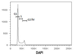
To study cell cycle analysis, C57BL/6 mouse bone marrow cells were stained with DRAQ5™ and acquired on a 633 nm laser-equipped flow cytometer. The above histogram shows DRAQ5™ detected with the 715 nm filter (PE/Cy7 channel). -

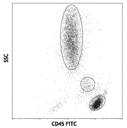
C57BL/6 mouse bone marrow cells were stained with CD45 FITC and DRAQ5™ (1:1000). Cells were acquired on a 633nm laser-equipped flow cytometer. -

HeLa cells were fixed with 1% paraformaldehyde (PFA) and blocked with 5% fetal bovine serum for 30 minutes at room temperature. Then the cells were stained with 10 µg/ml of EGFR (clone AY13) Brilliant Violet 421™ (blue) for three hours at room temperature. Nuclei were counterstained with 1:250 dilution of DRAQ5 (red). The image was captured with a 20 x objective. -

C57BL/6 mouse frozen spleen section was fixed with 4% paraformaldehyde (PFA) for 10 minutes at room temperature and blocked with 5% FBS plus 5% rat serum for 1 hour at room temperature. Then the section was stained with 2.5 µg/mL of CD3 (clone 17A2) Alexa Fluor® 647 (green), and 2.5 µg/mL of B220 (clone RA3-6B2) Brilliant Violet 421™ (blue) overnight at 4°C. Nuclei were counterstained with 1:250 of DRAQ5 (red). The image was captured by 10X objective. -

Human paraffin-embedded placenta tissue slice was prepared with a standard protocol of deparaffinization and rehydration. Antigen retrieval was done with Tris-Buffered Saline 1X (1.0 M, pH 7.4) at 95°C for 40 minutes. Tissue was washed with PBS/0.05% Tween 20 twice for five minutes and blocked with 5% FBS and 0.2% gelatin for 30 minutes. Then, the tissue was stained with 10 µg/mL of anti-human CD39 (clone A1) Brilliant Violet 421™ (blue) at 4°C overnight. Nuclei were counter stained with 1:250 dilution of DRAQ5™ (red). The image was captured with a 10X objective.
| Cat # | Size | Price | Quantity Check Availability | Save | ||
|---|---|---|---|---|---|---|
| 424101 | 50 µL | 175€ | ||||
DRAQ5™ is a far-red emitting, anthraquinone compound that stains nuclei in live cells. It is permeant to live cells and thus can be used for cell cycle analysis and/or staining of nucleated cells. It is optimally excited at 568 nm, 633 nm, and 647nm and can be detected using 695LP, 715LP, and 780LP filters. DRAQ5™ can also be used in cell imaging and is a good replacement for DAPI, as it does not get excited by UV or Violet lasers and is sub-optimally excited by the 488 nm laser. DRAQ5™ can be combined with FITC, PE, and other UV or violet excitable dyes for multi-color analysis.
Product DetailsProduct Details
- Preparation
- DRAQ5™ is supplied at 50 µL per vial (5 mM).
- Concentration
- Lot-specific (to obtain lot-specific concentration and expiration, please enter the lot number in our Certificate of Analysis online tool.)
- Storage & Handling
- DRAQ5™ should be stored between 2°C and 8°C upon receipt.
- Application
-
FC, ICFC - Quality tested
ICC, IHC-F, IHC-P - Verified - Recommended Usage
-
DRAQ5™ is a trademark of Biostatus Limited.
- Application Notes
-
DRAQ5™ is a cell-permeant DNA binding anthraquinone dye that intercalates between A-T bases of dsDNA. Due to its cell permeability, this dye is useful for assessing DNA content and cell cycle but is not suitable to be used as a viability dye. In microscopy applications, DRAQ5™ is also useful as a nuclear counterstain for both live and fixed specimens. It is a far-red emitting dye with optimal excitation/ emission peaks of 633nm/ 695nm, respectively, (when intercalated into DNA) that can be detected in the Alexa Fluor® 647, APC and Alexa Fluor® 700 channels. For use in cell cycle analysis, the optimal concentration must be determined for each cell type. Also, due to the wide emission spectrum and the brightness of DRAQ5™ off the 633nm red laser and the dim emission intensity of the APC-Cy7 or APC-Fire™ 750 fluorophores, flow cytometric panels should be optimized to determine that including DRAQ5™ in a panel is compatible with the use of the APC-Cy7 or APC-Fire™ 750 channel as well.
Protocol for DNA staining using DRAQ5™:
1. Perform surface staining following protocol of choice.
2. Wash cells twice with phosphate buffered saline (PBS). Sodium azide interferes with DRAQ5™ staining, thus it is recommended to stain in PBS (without calcium, magnesium, or sodium azide) or culture medium.
3. Dilute DRAQ5™ to required concentration. We recommend titrating the reagent to determine optimal concentration for cells of interest. Good results in flow cytometry and microscopy applications have been observed using a 1:200 to 1:1000 dilution.
4. Incubate at room temperature, preventing exposure to light, for 10-15 minutes. Subsequent washing can help make the cell cycle profile more distinct but is not absolutely necessary since DRAQ5™ is a fluorogenic reagent.
5. Analyze cells on a cytometer equipped with the 633 red laser. If performing cell cycle analysis, detection in the Alexa 700 channel with a 680LP or 715LP filter might help with resolution of the emission peaks. - Additional Product Notes
-
View more applications data for this product in our Scientific Poster Library.
- Product Citations
-
Antigen Details
- Biology Area
- Cell Biology, Cell Cycle/DNA Replication, Cell Motility/Cytoskeleton/Structure, Cell Proliferation and Viability, Neuroscience
- Antigen References
-
1. Smith PJ, et al. 1999. J. Immunol. Methods. 229:131.
2. Smith PJ, et al. 2000. Cytometry. 40:280.
3. Yuan CM, et al. 2004. Cytometry B. Clin. Cytom. 58:47. - Gene ID
- NA


Follow Us