 Login / Register


- Clone
- 3D6C02 (See other available formats)
- Regulatory Status
- RUO
- Other Names
- Nuclear factor of kappa light chain enhancer in B cells-inhibitor alpha, IKBA
- Isotype
- Mouse IgG2b, κ
- Ave. Rating
- Submit a Review
- Product Citations
- publications

-

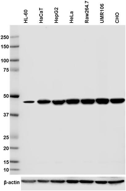
Western blot analysis of cell lysates from HeLa (Human), Raw264.7 (Mouse) and UMR106 (Rat) using IκB-α Mouse primary antibody and HRP Goat anti-Mouse secondary antibody (Cat. No. 405306). Direct-Blot™ HRP anti-β-actin (Cat. No. 643807) was used as a loading control. -

Total lysates (15 µg protein) from HepG2 (Human) and CHO (Hamster) were resolved by electrophoresis (4-20% Tris-glycine gel), transferred to nitrocellulose, and probed with 1:5000 (0.1 µg/ml) purified anti-IκB-α antibody, clone 3D6C02. Proteins were visualized using chemiluminescence detection by incubation with HRP Goat anti-Mouse secondary antibody (Cat. No. 405306, 1:3000 dilution). Direct-Blot™ HRP anti-β-actin antibody was used as a loading control (Cat. No. 643807, 1:8000 dilution). -

Hela cells were fixed with cold methanol at -20°C for 15 minutes and blocked with 5% FBS for 60 minutes. The cells were then intracellularly stained with (A) 2 µg/ml anti-mouse IgG2b, κ or (B) 2 µg/ml IκB-α antibody (clone 3D6C02), (C) 0.5 µg/ml IκB-α antibody (clone 3D6C02), or (D) 0.2 µg/ml IκB-α antibody (clone 3D6C02), and incubated overnight at 4°C followed by 2µg/ml (1:250) Alexa Fluor® 594 (Red) goat anti-mouse IgG for one hour at room temperature. Nuclei were counterstained with DAPI (Blue). The image was captured with a 60X objective. Exposure times: (A) 1/30, (B) 1/50, (C) 1/50, (D) 1/50. -

HeLa cells were stained with purified anti-IκBα (clone 3D6C02) antibody, followed by staining with DyLight™ 488 conjugated goat anti-mouse IgG (green) antibody and Alexa Fluor® 594 conjugated phalloidin (red). Nuclei were stained with DAPI (blue).
| Cat # | Size | Price | Quantity Check Availability | Save | ||
|---|---|---|---|---|---|---|
| 662402 | 100 µg | 184€ | ||||
IκB-α is an inhibitory protein that binds to the NF-κB complex and retains this complex in the cytoplasm where it cannot activate transcription. Upon cellular stimulation, the IκB proteins are phosphorylated and degraded via the ubiquitin pathway allowing for nuclear translocation of NF-κB. The NF-κB complex can then bind DNA to activate gene expression of multiple proteins involved in inflammation, cellular proliferation, differentiation, and apoptosis.
Product DetailsProduct Details
- Verified Reactivity
- Human, Mouse, Rat, Hamster
- Antibody Type
- Monoclonal
- Host Species
- Mouse
- Immunogen
- Full length human IκB-α recombinant protein expressed in E. coli.
- Formulation
- Phosphate-buffered solution, pH 7.2, containing 0.09% sodium azide.
- Preparation
- The antibody was purified by affinity chromatography.
- Concentration
- 0.5 mg/ml
- Storage & Handling
- The antibody solution should be stored undiluted between 2°C and 8°C.
- Application
-
WB - Quality tested
ICC - Verified - Recommended Usage
-
Each lot of this antibody is quality control tested by Western blotting. For Western blotting, the suggested use of this reagent is 0.1 - 0.5 µg/ml (1:1000 - 1:5000 dilution). For immunocytochemistry, the suggested use is 0.5 - 1.0 µg/ml (1:500-1:1000 dilution). It is recommended that the reagent be titrated for optimal performance for each application.
- Application Notes
-
This antibody weakly reacts with mouse and rat. Based on in-house testing, clone 3D6C02 does not block clone 25/IkBa/MAD-3, and only partially blocks L35A5 also raised against I?B-a.
- Product Citations
-
- RRID
-
AB_2564258 (BioLegend Cat. No. 662402)
Antigen Details
- Structure
- Inhibitory protein of the NF-κB complex that contains multiple ankyrin repeats.
- Distribution
-
Cytoplasm
- Function
- Regulates transcriptional activity of NF-κB by binding the complex and trapping it in the cytoplasm. Serine phosphorylation of IκB targets it for destruction via the ubiquitination pathway, which allows the NF-κB complex to translocate to the nucleus.
- Interaction
- NFkB1, SUMO4, p53, ribosomal S6 kinase 1
- Biology Area
- Apoptosis/Tumor Suppressors/Cell Death, Cell Biology, Signal Transduction
- Molecular Family
- Tumor Suppressors
- Antigen References
-
1. Baeuerle PA. 1998. Cell 95:729.
2. Hoffmann A, et al. 2002. Science 298:1241.
3. Ito CY, et al. 1995. Genomics 29:490. - Gene ID
- 4792 View all products for this Gene ID
- UniProt
- View information about IkappaB-alpha on UniProt.org

Related FAQs
Other Formats
View All IκB-α Reagents Request Custom Conjugation| Description | Clone | Applications |
|---|---|---|
| Purified anti-IκB-α | 3D6C02 | WB,ICC |
| Brilliant Violet 421™ anti-IκB-α | 3D6C02 | ICC |
| PE anti-IκB-α | 3D6C02 | ICFC |
Customers Also Purchased
Compare Data Across All Formats
This data display is provided for general comparisons between formats.
Your actual data may vary due to variations in samples, target cells, instruments and their settings, staining conditions, and other factors.
If you need assistance with selecting the best format contact our expert technical support team.
-
Purified anti-IκB-α

Western blot analysis of cell lysates from HeLa (Human), Raw... 
HeLa cells were stained with purified anti-IκBα (clone 3D6C0... 
Hela cells were fixed with cold methanol at -20°C for 15 min... 
Total lysates (15 µg protein) from HepG2 (Human) and CHO (Ha... -
Brilliant Violet 421™ anti-IκB-α

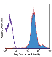
HeLa cells were fixed with cold 100% methanol for 10 minute... -
PE anti-IκB-α

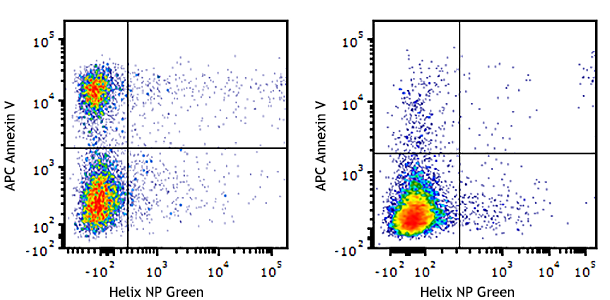
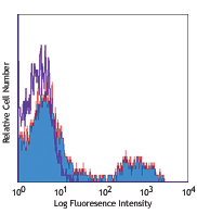
Human peripheral blood mononuclear cells were treated with (... 
Thioglycolate-elicited BALB/c mouse peritoneal macrophages w...


Follow Us