- Clone
- A18166G (See other available formats)
- Regulatory Status
- RUO
- Other Names
- Enolase 1; Alpha-Enolase; 2-Phospho-D-Glycerate Hydro-Lyase; Plasminogen-Binding Protein; Phosphopyruvate Hydratase
- Isotype
- Mouse IgG2b, κ
- Ave. Rating
- Submit a Review
- Product Citations
- publications

-

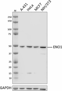
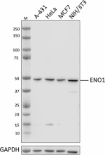
Whole cell extracts (15 µg total protein) from indicated cell lines were resolved by 4-12% Bis-Tris gel electrophoresis, transferred to a PVDF membrane, and probed with 0.05 µg/mL purified anti-ENO1 antibody (clone A18166G) overnight at 4°C. Proteins were visualized by chemiluminescence detection using HRP goat anti-mouse IgG antibody (Cat. No. 405306) at a 1:3000 dilution. Direct-Blot™ HRP anti-GAPDH antibody (Cat. No. 607904) was used as a loading control at a 1:50000 dilution (lower). Lane M: Molecular weight marker. -

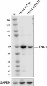
Whole cell extracts (15 µg protein) from HeLa cells transfected with non-targeting control siRNA (siCon) or siRNA targeting ENO1 (siENO1) were resolved on a 4-12% Bis-Tris gel, transferred to PVDF and probed with 1.0 µg /mL (1:500 dilution) purified anti-ENO1 antibody (clone A18166G), overnight at 4°C. Proteins were visualized by chemiluminescence detection using HRP goat anti-mouse IgG antibody (Cat. No. 405306) at a 1:3000 dilution. Direct-Blot™ HRP anti-GAPDH antibody (Cat. No. 607904) was used as a loading control at a 1:50000 dilution (lower). Lane M: Molecular Weight marker. -

HeLa cells were fixed with ice-cold methanol for 10 minutes and blocked with 5% FBS for 60 minutes. Cells were then intracellularly stained with 5.0 µg /mL purified mouse IgG2b, κ isotype control antibody (Cat. No. 400302) (panel A) or 1.0 µg/mL (1:500 dilution) purified anti-ENO1 antibody (clone A18166G) (panel B) overnight at 4°C, followed by incubation with Alexa Fluor® 594 goat anti-mouse IgG antibody (Cat. No. 405326) at a 1:200 dilution. Nuclei were counterstained with DAPI and the image was captured with a 60X objective. -

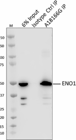
Whole cell extracts (250 µg total protein) prepared from HeLa cells were immunoprecipitated overnight with 2.5 µg of purified mouse IgG2b, κ isotype control antibody (Cat. No. 400302) or purified anti-ENO1 antibody (clone A18166G). The resulting IP fractions and whole cell extract input (6%) were resolved by 4-12% Bis-Tris gel electrophoresis, transferred to a PVDF membrane and probed with a rabbit control antibody against a separate epitope of ENO1. Lane M: Molecular weight marker.
| Cat # | Size | Price | Quantity Check Availability | Save | ||
|---|---|---|---|---|---|---|
| 945501 | 25 µg | 95€ | ||||
| 945502 | 100 µg | 235€ | ||||
Enolase is highly conserved, multifunctional protein. In the cytoplasm, ENO1 acts as a metabolic enzyme and catalyzes 2-phosphoglycerate to phosphoenolpyruvate in the last step of the glycolytic cycle.
Despite the lack of signal sequence in the ENO1 gene, ENO1 is found to be expressed on the surface of several hematopoietic cells though the transport mechanism is still unknown. Several of these cells include monocytes, T cells and B cells, and endothelial cells. On the cell surface, ENO1 acts as a plasminogen receptor and concentrates plasmin proteolytic activity at the pericellular area and protects plasmin from α2-antiplasmin inhibitors.
In cancer cells, ENO1 promotes what is known as the Warburg effect, a modified cellular metabolism in which the tumor cells uptake excessive glucose and undergo glycolysis to produce lactate in both hypoxic conditions and normal oxygen levels. ENO1 also promotes tumor invasion through plasminogen activation and extracellular matrix degradation.
Enolase can also be found in neurons and peripheral neuroendocrine cells. This neuron-specific enolase exists as ENO2 homodimer or an ENO1 and ENO2 heterodimer. Raised serum levels of these neuron-specific enolase have been found in all stages of neuroblastoma and it has been proposed that the plasmin formation caused by ENO1 enhances neuritogenesis.
The ENO1 gene can also be alternatively spliced and produce a 37 kD protein named c-myc promoter-binding protein, or MBP-1. MBP-1 is localized to the nucleus and binds to the c-myc P2 promoter, preventing the transcription of proto-oncogenes.
Product Details
- Verified Reactivity
- Human, Mouse
- Antibody Type
- Monoclonal
- Host Species
- Mouse
- Immunogen
- Recombinant human ENO1 protein
- Formulation
- Phosphate-buffered solution, pH 7.2, containing 0.09% sodium azide
- Preparation
- The antibody was purified by affinity chromatography.
- Concentration
- 0.5 mg/mL
- Storage & Handling
- The antibody solution should be stored undiluted between 2°C and 8°C.
- Application
-
WB - Quality tested
KO/KD-WB, ICC, IP - Verified - Recommended Usage
-
Each lot of this antibody is quality control tested by western blotting. For western blotting, the suggested use of this reagent is 0.05 - 1.0 µg/mL. For immunocytochemistry, a concentration range of 1.0 - 5.0 μg/mL is recommended. For immunoprecipitation, the suggested use of this reagent is 2.5 µg/test. It is recommended that the reagent be titrated for optimal performance for each application.
- Application Notes
-
When tested for ICC using 4%-PFA fixed HeLa cells permeabilized with methanol or Triton X-100, high non-specific staining in the nucleus was observed. We therefore do not recommend these fix-perm methods for this clone.
- RRID
-
AB_2892513 (BioLegend Cat. No. 945501)
AB_2892513 (BioLegend Cat. No. 945502)
Antigen Details
- Structure
- ENO1 is a 434 amino acid protein with a predicted molecular weight of 47 kD.
- Distribution
-
Cytosolic/Ubiquitously expressed
- Function
- Glycolytic enzyme
- Biology Area
- Cell Biology
- Antigen References
-
- Capello M, et al. 2011. FEBS J. 278:1064-74
- Capello M, et al. 2016. Oncotarget. 7:5598-5612
- Cappello P, et al. 2017. Front Biosci (Landmark Ed). 22:944-959
- Díaz-Ramos A, et al. 2012. J Biomed Biotechnol. 2012:156795
- Didiasova M, et al. 2019. Front Cell Dev Biol. 7:61
- Isgrò MA, et al. 2015. Adv Exp Med Biol. 867:125-43.
- Liberti MV and Locasale JW. 2016. Trends Biochem Sci. 41:211-218.
- Gene ID
- 2023 View all products for this Gene ID
- UniProt
- View information about ENO1 on UniProt.org
Related Pages & Pathways
Pages
Related FAQs
Other Formats
View All ENO1 Reagents Request Custom Conjugation| Description | Clone | Applications |
|---|---|---|
| Purified anti-ENO1 | A18166G | WB,KO/KD-WB,ICC,IP |
Compare Data Across All Formats
This data display is provided for general comparisons between formats.
Your actual data may vary due to variations in samples, target cells, instruments and their settings, staining conditions, and other factors.
If you need assistance with selecting the best format contact our expert technical support team.
 Login / Register


Follow Us