 Login / Register


- Clone
- RMP1-14 (See other available formats)
- Regulatory Status
- RUO
- Other Names
- Programmed Death-1, CD279, PD 1, PDCD1
- Isotype
- Rat IgG2a, κ
- Ave. Rating
- Submit a Review
- Product Citations
- publications

-

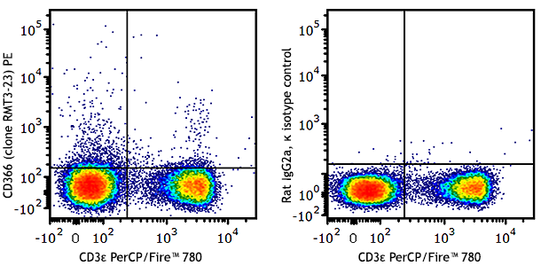
Con A-stimulated (3 days) C57BL/6 splenocytes were stained with CD279 (clone RMP1-14) PE (filled histogram) or rat IgG2a, κ PE isotype control (open histogram).
| Cat # | Size | Price | Quantity Check Availability | Save | ||
|---|---|---|---|---|---|---|
| 114117 | 50 µg | 108€ | ||||
| 114118 | 200 µg | 302€ | ||||
CD279 is a 50-55 kD immunoglobulin superfamily member, also known as programmed death-1 (PD-1). PD-1 is expressed on a subset of CD4-CD8- thymocytes, and on activated T and B cells. PD-1 is thought to be involved in lymphocyte clonal selection and peripheral tolerance. The PD-1 ligands, PD-L1 (also known as B7-H1) and PD-L2 (B7-DC), are members of the B7 immunoglobulin superfamily. The RMP1-14 antibody has been reported to block the binding of PD-1 to its ligands (B7-H1 and B7-DC) and to inhibit T cell proliferation and cytokine production costimulated by macrophages (but not by dendritic cells and B cells).
Product DetailsProduct Details
- Verified Reactivity
- Mouse
- Antibody Type
- Monoclonal
- Host Species
- Rat
- Immunogen
- Mouse PD-1 transfected BHK cells
- Formulation
- Phosphate-buffered solution, pH 7.2, containing 0.09% sodium azide.
- Preparation
- The antibody was purified by affinity chromatography and conjugated with PE under optimal conditions.
- Concentration
- 0.2 mg/ml
- Storage & Handling
- The antibody solution should be stored undiluted between 2°C and 8°C, and protected from prolonged exposure to light. Do not freeze.
- Application
-
FC - Quality tested
- Recommended Usage
-
Each lot of this antibody is quality control tested by immunofluorescent staining with flow cytometric analysis. For flow cytometric staining, the suggested use of this reagent is ≤1.0 µg per million cells in 100 µl volume. It is recommended that the reagent be titrated for optimal performance for each application.
- Excitation Laser
-
Blue Laser (488 nm)
Green Laser (532 nm)/Yellow-Green Laser (561 nm)
- Application Notes
-
Additional reported applications (for relevant formats of this clone) include: immunohistochemical staining of acetone-fixed frozen sections1, Western blotting2,6, inhibition of macrophages (but not B cells and dendritic cells) costimulated T cell proliferation3,5, and blocking the binding of PD-1 to B7-H1 or B7-DC3,4,8. The LEAF™ purified antibody (Endotoxin <0.1 EU/µg, Azide-Free, 0.2 µm filtered) is recommended for functional assays (Cat. No. 114108). For highly sensitive assays, we recommend Ultra-LEAF™ purified antibody (Cat. No. 114110) with a lower endotoxin limit than standard LEAF™ purified antibodies.
- Application References
-
- Kanai, T., et al. 2003. J. Immunol. 171:4156. (FC, IHC)
- Personal Communication (WB)
- Yamazaki, T., et al. 2005. J. Immunol. 175:1586. (Costim)
- Matsumoto, K., et al. 2004. J. Immunol. 172:2530. (Block)
- Terrazas, L. I., et al. 2005. Intl. J. Parasitology. 35:1349. (Costim)
- Meng, Q., et al. 2006. Invest Opthalmol Vis Sci. 47:444. (FC, WB) PubMed
- Tsang JY, et al. 2011. Int Immunopharmacol. 11:604. PubMed
- Takamura S, et al. 2010. J. Immunol. 184:4696. PubMed (Block)
- Product Citations
-
- RRID
-
AB_2566725 (BioLegend Cat. No. 114117)
AB_2566726 (BioLegend Cat. No. 114118)
Antigen Details
- Structure
- Ig superfamily, 50-55 kD
- Distribution
-
Subset of double negative thymocytes, activated T and B cells
- Function
- Lymphocyte clonal selection, peripheral tolerance
- Ligand/Receptor
- PD-L1 (B7-H1), PD-L2
- Cell Type
- B cells, T cells, Thymocytes, Tregs
- Biology Area
- Apoptosis/Tumor Suppressors/Cell Death, Cancer Biomarkers, Cell Biology, Immunology, Inhibitory Molecules
- Molecular Family
- CD Molecules, Immune Checkpoint Receptors
- Antigen References
-
1. Barclay A, et al. 1997. The Leukocyte Antigen FactsBook, Academic Press.
2. Agata Y, et al. 1996. Int. Immunol. 8:765.
3. Nishimura H, et al. 2001. Science 291:319.
4. Ishida Y, et al. 1992. EMBO J. 11:3887. - Gene ID
- 18566 View all products for this Gene ID
- UniProt
- View information about CD279 on UniProt.org
Related Pages & Pathways
Pathways


Related FAQs
- What type of PE do you use in your conjugates?
- We use R-PE in our conjugates.
Other Formats
View All CD279 Reagents Request Custom Conjugation| Description | Clone | Applications |
|---|---|---|
| Purified anti-mouse CD279 (PD-1) | RMP1-14 | FC,IHC,WB,FA |
| LEAF™ Purified anti-mouse CD279 (PD-1) | RMP1-14 | FC, IHC, WB, FA |
| Ultra-LEAF™ Purified anti-mouse CD279 (PD-1) | RMP1-14 | FC,IHC,WB,FA |
| GoInVivo™ Purified anti-mouse CD279 (PD-1) | RMP1-14 | FC,IHC,WB,FA |
| PE anti-mouse CD279 (PD-1) | RMP1-14 | FC |
Customers Also Purchased
Compare Data Across All Formats
This data display is provided for general comparisons between formats.
Your actual data may vary due to variations in samples, target cells, instruments and their settings, staining conditions, and other factors.
If you need assistance with selecting the best format contact our expert technical support team.
-
Purified anti-mouse CD279 (PD-1)

Con A-stimulated C57BL/6 splenocytes stained with purified R... -
LEAF™ Purified anti-mouse CD279 (PD-1)

Con A-stimulated C57BL/6 splenocytes stained with LEAF™ puri... -
Ultra-LEAF™ Purified anti-mouse CD279 (PD-1)

Con A-stimulated (3 days) C57BL/6 splenocytes were stained w... -
GoInVivo™ Purified anti-mouse CD279 (PD-1)

Anti-mouse PD-1 inhibits the binding of mouse PD-1 to PD-L1.... -
PE anti-mouse CD279 (PD-1)

Con A-stimulated (3 days) C57BL/6 splenocytes were stained w...


Follow Us