 Login / Register


- Clone
- A18002D (See other available formats)
- Regulatory Status
- RUO
- Other Names
- Lsk, p56lck, LCK Proto-Oncogene, Src Family Tyrosine Kinase, Tyrosine-protein kinase LCK, Leukocyte C-terminal Src kinase, LSK, Lymphocyte cell-specific protein-tyrosine kinase, T cell-specific protein-tyrosine kinase
- Isotype
- Mouse IgG1, κ
- Ave. Rating
- Submit a Review
- Product Citations
- publications

-

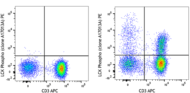
Human peripheral blood lymphocytes were treated with (left), or without (right) hydrogen peroxide for 5 minutes, fixed with Fixation Buffer (Cat. No. 420801), permeabilized with Intracellular Staining Permeabilization Wash Buffer (Cat. No. 421002), then surfaced stained with CD3 APC and intracellularly stained with anti-Lck Phospho (Tyr394) (clone A18002D) PE.
| Cat # | Size | Price | Quantity Check Availability | Save | ||
|---|---|---|---|---|---|---|
| 933103 | 25 tests | 117€ | ||||
| 933104 | 100 tests | 298€ | ||||
The Src family tyrosine kinase p56Lck (Lck) is a non-receptor tyrosine kinase that plays a critical role in T cell selection and maturation within the thymus, and also in the function of mature T cells. Lck, which is constitutively bound to cytosolic domains of CD4 and CD8 surface receptors, plays an essential role in T cell receptor (TCR) signaling. Engagement of the TCR with peptide antigen-loaded MHC complexes results in the recruitment of CD4- and CD8-bound Lck to the TCR/CD3 signaling complex. Lck then transphosphorylates TCR-gamma chains and CD3 subunits, thereby activating the TCR/CD3 signaling pathway and leading to the recruitment and subsequent phosphorylation of Zap70 by Lck. Lck also plays an important role in interleukin-2 signaling that regulates the T cell proliferative response. Phosphorylation of Lck by CSK at tyrosine 505 negatively regulates the kinase, and is proposed to generate a closed, inactive conformation of the protein. Conversely, phosphorylation at tyrosine 394 in the activation loop of Lck greatly stimulates enzymatic activity. In many types of cancer, Lck is a proliferative and anti-apoptotic driver, and has been proposed as a target for therapeutic intervention.
Product DetailsProduct Details
- Verified Reactivity
- Human, Mouse
- Antibody Type
- Monoclonal
- Host Species
- Mouse
- Immunogen
- Synthetic peptide corresponding to human Src phosphorylated at tyrosine 419
- Formulation
- Phosphate-buffered solution, pH 7.2, containing 0.09% sodium azide and BSA (origin USA)
- Preparation
- The antibody was purified by affinity chromatography and conjugated with PE under optimal conditions.
- Concentration
- Lot-specific (to obtain lot-specific concentration and expiration, please enter the lot number in our Certificate of Analysis online tool.)
- Storage & Handling
- The antibody solution should be stored undiluted between 2°C and 8°C, and protected from prolonged exposure to light. Do not freeze.
- Application
-
ICFC - Quality tested
- Recommended Usage
-
Each lot of this antibody is quality control tested by intracellular immunofluorescent staining with flow cytometric analysis. For flow cytometric staining, the suggested use of this reagent is 5 µL per million cells in 100 µL staining volume or 5 µL per 100 µL of whole blood.
- Excitation Laser
-
Blue Laser (488 nm)
Green Laser (532 nm)/Yellow-Green Laser (561 nm)
- Application Notes
-
This clone has not been tested for IP.
This clone may recognize other Src family members.
This clone was ICC tested on PFA-fixed cells using both methanol and Triton X-100 permeabilization methods. Both permeabilazation methods were compatible with staining. Triton X-100 permeabilization produces stronger staining.
During ICC product development testing, this clone produced strong nuclear staining in a small subpopulation (<1%) of both unstimulated and stimulated cells. The source of this staining is not clear but we do not believe it is Lck-dependent.
This clone was developed using a synthetic peptide corresponding to human Src phosphorylated at tyrosine 419, which displays high sequence homology with the region surrounding Lck tyrosine 394. To confirm specificity to Lck Phospho (Tyr394), the cell line J.Cam1.6 was used. This cell line is a clonal derivative of Jurkat cells that harbors a truncated LCK allele with defective protein expression (Straus, et al. 1992. Cell. 70:585). - Product Citations
-
- RRID
-
AB_2820203 (BioLegend Cat. No. 933103)
AB_2820204 (BioLegend Cat. No. 933104)
Antigen Details
- Structure
- Lck is a 509 amino acid protein with a predicted molecular weight of 58 kD
- Distribution
-
Plasma-membrane associated/T cells
- Function
- Protein Tyrosine Kinase
- Cell Type
- T cells
- Biology Area
- Cell Biology, Signal Transduction
- Molecular Family
- Protein Kinases/Phosphatase
- Antigen References
-
- Phillipsen L, et al. 2017. Sci Signal. 10:eaaf4736.
- Moogk D, et al. 2016. J Immunol. 197:644.
- Klammt C, et al. 2015. Nat Immunol. 16:961.
- Cancer Genome Atlas Network. 2015. Cell. 161:1681.
- Casas J, et al. 2014. Nat Commun. 5:5624
- McNeill L, et al. 2007. Immunnity. 27:425.
- Lefebvre DC, et al. 2003. Biochim Biophys Acta. 1650:40.
- Kabouridis PS. et al. 2003. Biochem J. 371:907.
- Abraham N, et al. 1990. Mol. Cell. Biol. 10:5197.
- Gene ID
- 3932 View all products for this Gene ID
- UniProt
- View information about Lck Phospho Tyr394 on UniProt.org
Related Pages & Pathways
Pathways


Related FAQs
- What type of PE do you use in your conjugates?
- We use R-PE in our conjugates.
Other Formats
View All Lck Phospho (Tyr394) Reagents Request Custom Conjugation| Description | Clone | Applications |
|---|---|---|
| Purified anti-Lck Phospho (Tyr394) | A18002D | WB,ICC,ICFC |
| PE anti-Lck Phospho (Tyr394) | A18002D | ICFC |
Customers Also Purchased
Compare Data Across All Formats
This data display is provided for general comparisons between formats.
Your actual data may vary due to variations in samples, target cells, instruments and their settings, staining conditions, and other factors.
If you need assistance with selecting the best format contact our expert technical support team.
-
Purified anti-Lck Phospho (Tyr394)

Whole cell extracts (15 µg protein) from serum starved Jurka... 
Whole cell extracts (15 µg protein) from serum starved EL4 c... 
Whole cell extracts (15 µg protein) from Jurkat and J.Cam1.6... 
Jurkat cells untreated (panel A) or treated (panel B) with 5... 
Human peripheral blood lymphocytes were treated with (left),... -
PE anti-Lck Phospho (Tyr394)

Human peripheral blood lymphocytes were treated with (left),...


Follow Us