 Login / Register


- Clone
- L161 (See other available formats)
- Regulatory Status
- RUO
- Workshop
- V T-CD01.18
- Other Names
- R7, M241, BDCA-1
- Isotype
- Mouse IgG1, κ
- Ave. Rating
- Submit a Review
- Product Citations
- publications

-

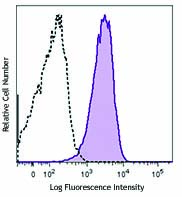
Human peripheral blood lymphocytes were stained with CD19 FITC and CD1c (clone L161) Brilliant Violet 650™ (left) or, Mouse IgG1, κ Brilliant Violet 650™ isotype control (right).
| Cat # | Size | Price | Quantity Check Availability | Save | ||
|---|---|---|---|---|---|---|
| 331541 | 25 tests | 172€ | ||||
| 331542 | 100 tests | 348€ | ||||
CD1c, also known as R7 or M241, is a 43 kD member of the five CD1 antigens (CD1a-e) in humans. The CD1 molecules are type I glycoprotein with structural similarities to MHC class I and are non-covalently associated with β2-microglobulin, belonging to the Ig superfamily. CD1c is expressed on cortical thymocytes, Langerhans cells, dendritic cells, and a subset of B cells. It has been reported that CD1c is also expressed on mature T cells in a tightly regulated manner. CD1c is involved in antigen-presentation of glycolipids. It may also act in T cells as an immune regulatory molecule.
Product DetailsProduct Details
- Verified Reactivity
- Human
- Reported Reactivity
- African Green, Baboon, Cynomolgus, Rhesus
- Antibody Type
- Monoclonal
- Host Species
- Mouse
- Formulation
- Phosphate-buffered solution, pH 7.2, containing 0.09% sodium azide and BSA (origin USA).
- Preparation
- The antibody was purified by affinity chromatography and conjugated with Brilliant Violet 650™ under optimal conditions.
- Concentration
- Lot-specific (to obtain lot-specific concentration and expiration, please enter the lot number in our Certificate of Analysis online tool.)
- Storage & Handling
- The antibody solution should be stored undiluted between 2°C and 8°C, and protected from prolonged exposure to light. Do not freeze.
- Application
-
FC - Quality tested
- Recommended Usage
-
Each lot of this antibody is quality control tested by immunofluorescent staining with flow cytometric analysis. For flow cytometric staining, the suggested use of this reagent is 5 µl per million cells in 100 µl staining volume or 5 µl per 100 µl of whole blood.
Brilliant Violet 650™ excites at 405 nm and emits at 645 nm. The bandpass filter 660/20 nm is recommended for detection, although filter optimization may be required depending on other fluorophores used. Be sure to verify that your cytometer configuration and software setup are appropriate for detecting this channel. Refer to your instrument manual or manufacturer for support. Brilliant Violet 650™ is a trademark of Sirigen Group Ltd.
Learn more about Brilliant Violet™.
This product is subject to proprietary rights of Sirigen Inc. and is made and sold under license from Sirigen Inc. The purchase of this product conveys to the buyer a non-transferable right to use the purchased product for research purposes only. This product may not be resold or incorporated in any manner into another product for resale. Any use for therapeutics or diagnostics is strictly prohibited. This product is covered by U.S. Patent(s), pending patent applications and foreign equivalents. - Excitation Laser
-
Violet Laser (405 nm)
- Application Notes
-
Additional reported applications (for the relevant formats) include: immunohistochemical staining on frozen tissue4, 5, formalin-fixed paraffin-embedded immunohistochemical staining6, and spatial biology (IBEX)7,8.
- Application References
-
- del C Salamone M, et al. 2001. J Leukoc Biol. 70:567.
- de Fraissinette A, et al. 1988. Exp Hematol. 16:764.
- Li D, et al. 2012. J Exp Med. 209:109. PubMed
- Xu C, et al. 2010. Am J Hematol. 85:539 (IHC-F)
- Gerlini G, et al. 2001. J Invest Dermatol. 117:576 (IHC-F)
- Poposki J, et al. 2016. Clin Exp Allergy 45:384 (IHC-P) PubMed
- Radtke AJ, et al. 2020. Proc Natl Acad Sci USA. 117:33455-33465. (SB) PubMed
- Radtke AJ, et al. 2022. Nat Protoc. 17:378-401. (SB) PubMed
- Product Citations
-
- RRID
-
AB_2800865 (BioLegend Cat. No. 331541)
AB_2800866 (BioLegend Cat. No. 331542)
Antigen Details
- Structure
- 43 kD, Ig superfamily, MHC I-like molecule, associates with β2-microglobulin
- Distribution
-
B cell subset, cortical thymocytes, dendritic cells, and Langerhans cells
- Function
- Presents lipid antigen to CD1c-restricted T cells
- Ligand/Receptor
- CD1c-restricted TCR
- Cell Type
- B cells, Dendritic cells, Langerhans cells, Thymocytes
- Biology Area
- Immunology
- Molecular Family
- CD Molecules
- Antigen References
-
1. Fainboim LM and del C. Salamone. 2002. J. Biol. Reg. Homeos. Ag. 16:125.
2. M. del Salamone C, et al. 2001. J. Leukocyte Biol. 70:567.
3. Zola H, et al. Eds. 2007. Leukocyte and Stromal Cell Molecules:The CD Markers. P42. - Gene ID
- 911 View all products for this Gene ID
- UniProt
- View information about CD1c on UniProt.org


Other Formats
View All CD1c Reagents Request Custom ConjugationCustomers Also Purchased
Compare Data Across All Formats
This data display is provided for general comparisons between formats.
Your actual data may vary due to variations in samples, target cells, instruments and their settings, staining conditions, and other factors.
If you need assistance with selecting the best format contact our expert technical support team.
-
PerCP anti-human CD1c

Human peripheral blood lymphocytes stained with L161 PerCP -
Purified anti-human CD1c

Human peripheral blood lymphocytes stained with purified L16... -
Biotin anti-human CD1c

Human peripheral blood lymphocytes stained with biotinylated... -
PE anti-human CD1c

Human peripheral blood lymphocytes stained with L161 PE 
Confocal image of human lymph node sample acquired using the... -
Pacific Blue™ anti-human CD1c

Human T lymphoblastic leukemia cell line, Molt-4, stained wi... -
Alexa Fluor® 647 anti-human CD1c

Human T lymphoblastic leukemia cell line, Molt-4, stained wi... -
PerCP/Cyanine5.5 anti-human CD1c

Human peripheral blood lymphocytes were stained with CD19 AP... -
Brilliant Violet 421™ anti-human CD1c

Human peripheral blood lymphocytes were stained with CD19 AP... 
-
PE/Cyanine7 anti-human CD1c

Human T lymphoblastic leukemia cell line Molt-4 stained with... -
FITC anti-human CD1c

Human T lymphoblastic cell line, Molt-4, stained with L161 F... -
APC/Cyanine7 anti-human CD1c

Human peripheral blood lymphocytes stained with CD19 FITC an... -
APC anti-human CD1c

Human peripheral blood lymphocytes were stained with CD19 FI... 
-
Alexa Fluor® 488 anti-human CD1c

Human peripheral blood lymphocytes were stained with CD19 AP... 
-
Alexa Fluor® 700 anti-human CD1c

Human peripheral blood lymphocytes were stained with CD19 PE... 
-
PE/Dazzle™ 594 anti-human CD1c

Human peripheral blood lymphocytes were stained with CD19 AP... 
-
Brilliant Violet 510™ anti-human CD1c

Human peripheral blood lymphocytes were stained with CD19 FI... 
-
Brilliant Violet 605™ anti-human CD1c

Human peripheral blood lymphocytes were stained with CD19 FI... 
-
Brilliant Violet 711™ anti-human CD1c

Human peripheral blood lymphocytes were stained with CD19 FI... 
-
TotalSeq™-A0160 anti-human CD1c
-
Brilliant Violet 650™ anti-human CD1c

Human peripheral blood lymphocytes were stained with CD19 FI... -
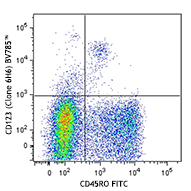
Brilliant Violet 785™ anti-human CD1c

Human peripheral blood lymphocytes were stained with CD19 FI... -
APC/Fire™ 750 anti-human CD1c

Human peripheral blood lymphocytes were stained with CD19 PE... -
TotalSeq™-C0160 anti-human CD1c
-
TotalSeq™-B0160 anti-human CD1c
-
TotalSeq™-D0160 anti-human CD1c
-
PE/Cyanine5 anti-human CD1c

Human peripheral blood lymphocytes were stained with HIB19 F... -
Spark Red™ 718 anti-human CD1c (Flexi-Fluor™)


Follow Us