 Login / Register


- Clone
- GL-1 (See other available formats)
- Regulatory Status
- RUO
- Other Names
- B7-2, B70, Ly-58
- Isotype
- Rat IgG2a, κ
- Ave. Rating
- Submit a Review
- Product Citations
- publications

-

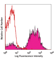
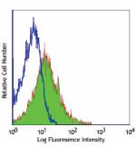
LPS-stimulated (3 days) C57BL/6 mouse splenocytes stained with GL-1 APC
| Cat # | Size | Price | Quantity Check Availability | Save | ||
|---|---|---|---|---|---|---|
| 105011 | 25 µg | 77€ | ||||
| 105012 | 100 µg | 227€ | ||||
CD86 is an 80 kD immunoglobulin superfamily member also known as B7-2, B70, and Ly-58. CD86 is expressed on activated B and T cells, macrophages, dendritic cells, and astrocytes. CD86, along with CD80, is a ligand of CD28 and CD152 (CTLA-4). CD86 is expressed earlier in the immune response than CD80. CD86 has also been shown to be involved in immunoglobulin class-switching and triggering of NK cell-mediated cytotoxicity. CD86 binds to CD28 to transduce co-stimulatory signals for T cell activation, proliferation, and cytokine production. CD86 can also bind to CD152, also known as CTLA-4, to deliver an inhibitory signal to T cells.
Product DetailsProduct Details
- Verified Reactivity
- Mouse
- Antibody Type
- Monoclonal
- Host Species
- Rat
- Immunogen
- LPS-activated CBA/Ca mouse splenic B cells
- Formulation
- Phosphate-buffered solution, pH 7.2, containing 0.09% sodium azide.
- Preparation
- The antibody was purified by affinity chromatography, and conjugated with APC under optimal conditions.
- Concentration
- 0.2 mg/ml
- Storage & Handling
- The antibody solution should be stored undiluted between 2°C and 8°C, and protected from prolonged exposure to light. Do not freeze.
- Application
-
FC - Quality tested
- Recommended Usage
-
Each lot of this antibody is quality control tested by immunofluorescent staining with flow cytometric analysis. For flow cytometric staining, the suggested use of this reagent is ≤ 0.25 µg per 106 cells in 100 µl volume. It is recommended that the reagent be titrated for optimal performance for each application.
- Excitation Laser
-
Red Laser (633 nm)
- Application Notes
-
The GL-1 antibody can block the mixed lymphocyte reaction in vitro and has been shown to inhibit the priming of cytotoxic T lymphocytes in vivo (along with antibodies against B7-1). Additional reported applications (for the relevant formats) include: immunoprecipitation1, immunohistochemical staining of acetone-fixed frozen sections2,6, immunofluorescence microscopy, and in vivo and in vitro blocking of T cell responses1-6. GL-1 is not suitable for immunohistochemical staining of formalin-fixed paraffin sections. The Ultra-LEAF™ purified antibody (Endotoxin < 0.01 EU/µg, Azide-Free, 0.2 µm filtered) is recommended for functional assays (Cat. No. 105051-105056).
- Application References
-
- Hathcock KS, et al. 1993. Science 262:905. (Block, IP)
- Inaba KM, et al. 1994. J. Exp. Med. 180:1849. (Block, IHC)
- Hathcock KS, et al. 1994. J. Exp. Med. 180:631. (Block)
- Krummel MF, et al. 1995. J. Exp. Med. 182:459. (Block)
- Liu Y, et al. 1997. J. Exp. Med. 185:251. (Block)
- Herold KC, et al. 1997. J. Immunol. 158:984. (Block, IHC)
- Shih FF, et al. 2006. J. Immunol. 176:3438. (FC)
- Lawson BR, et al. 2007. J. Immunol. 178:5366.
- Turnquist HR, et al. 2007. J. Immunol. 178:7018.
- Klinger MB, et al. 2007. Am. J. Physiol. Requl. Integr. Comp. Physiol. 293:R677. PubMed
- de Verteuil DA, et al. 2014. J Immunol. 193:1121. PubMed
- Product Citations
-
- RRID
-
AB_493343 (BioLegend Cat. No. 105011)
AB_493342 (BioLegend Cat. No. 105012)
Antigen Details
- Structure
- Ig superfamily, 80 kD
- Distribution
-
B cells and T cells (upregulated upon activation), macrophages, dendritic cells, and astrocytes
- Function
- T cell costimulation, Ig class-switching, NK cell cytotoxicity
- Ligand/Receptor
- CD28, CD152 (CTLA-4)
- Cell Type
- Astrocytes, B cells, Dendritic cells, Macrophages, T cells, Tregs
- Biology Area
- Cell Biology, Costimulatory Molecules, Immunology, Neuroscience, Neuroscience Cell Markers
- Molecular Family
- CD Molecules, Immune Checkpoint Receptors
- Antigen References
-
1. Barclay A, et al. 1997. The Leukocyte Antigen FactsBook Academic Press.
2. Hathcock KS, et al. 1993. Science 262:905.
3. Freeman GJ, et al. 1993. Science 262:907.
4. Carreno BM, et al. 2002. Annu. Rev. Immunol. 20:29. - Gene ID
- 12524 View all products for this Gene ID
- UniProt
- View information about CD86 on UniProt.org
Related Pages & Pathways
Pathways


Other Formats
View All CD86 Reagents Request Custom ConjugationCustomers Also Purchased
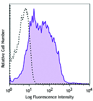
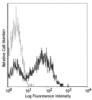
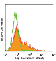
Compare Data Across All Formats
This data display is provided for general comparisons between formats.
Your actual data may vary due to variations in samples, target cells, instruments and their settings, staining conditions, and other factors.
If you need assistance with selecting the best format contact our expert technical support team.
-
Brilliant Violet 650™ anti-mouse CD86

LPS-stimulated (3 days) C57BL/6 mouse splenocytes were stain... -
Biotin anti-mouse CD86

C57BL/6 mouse splenocytes stained with biotinylated GL-1,fol... -
FITC anti-mouse CD86

LPS-stimulated (3 days) C57BL/6 mouse splenocytes stained wi... -
PE anti-mouse CD86

LPS (3-day) stimulated BALB/c mouse splenocytes stained with... -
Purified anti-mouse CD86

C57BL/6 mouse splenocytes stained with purified GL-1,followe... -
Brilliant Violet 605™ anti-mouse CD86

LPS-stimulated (3 days) C57BL/6 mouse splenocytes were stain... -
APC anti-mouse CD86

LPS-stimulated (3 days) C57BL/6 mouse splenocytes stained wi... -
PE/Cyanine7 anti-mouse CD86

C57BL/6 mouse splenocytes stained with GL-1 PE/Cyanine7 -
Alexa Fluor® 488 anti-mouse CD86

LPS-stimulated (3 days) C57BL/6 mouse splenocytes stained wi... -
Alexa Fluor® 647 anti-mouse CD86

LPS-stimulated (3 days) C57BL/6 mouse splenocytes stained wi... -
Pacific Blue™ anti-mouse CD86

LPS (3-day) stimulated C57BL/6 mouse splenocytes stained wit... -
PE/Cyanine5 anti-mouse CD86

C57BL/6 mouse splenocytes stained with GL-1 PE/Cyanine5 -
Alexa Fluor® 700 anti-mouse CD86

LPS-stimulated (3 days) C57BL/6 mouse splenocytes stained wi... -
PerCP/Cyanine5.5 anti-mouse CD86

LPS-stimulated (3 days) C57BL/6 mouse splenocytes were stain... -
PerCP anti-mouse CD86
-
APC/Cyanine7 anti-mouse CD86

LPS-stimulated (3 days) C57BL/6 splenocytes stained with GL-... -
Brilliant Violet 421™ anti-mouse CD86

LPS-stimulated (3 days) C57BL/6 mouse splenocytes stained wi... -
Brilliant Violet 510™ anti-mouse CD86

LPS stimulated C57BL/6 mouse splenocytes (3 days) were stain... -
PE/Dazzle™ 594 anti-mouse CD86

LPS-stimulated (three days) C57BL/6 mouse splenocytes were s... -
Brilliant Violet 785™ anti-mouse CD86

LPS stimulated C57BL/6 mouse splenocytes (3 days) were stain... -
APC/Fire™ 750 anti-mouse CD86

LPS-stimulated (3 days) C57BL/6 mouse splenocytes were stain... -
TotalSeq™-A0200 anti-mouse CD86
-
TotalSeq™-B0200 anti-mouse CD86
-
Ultra-LEAF™ Purified anti-mouse CD86
C57BL/6 mouse splenocytes stained with Ultra-LEAF™ purified ... -
TotalSeq™-C0200 anti-mouse CD86


Follow Us