Brilliant Violet 421™ Streptavidin

Pricing & Availability
Regulatory Status
RUO
Other Names
SAv-Brilliant Violet 421™
Ave. Rating
Submit a Review
Product Citations
publications
SAV_BV421_071911
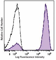
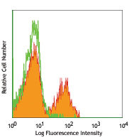
Human peripheral blood lymphocytes were stained with biotinylated CD3 (filled histogram) or mouse IgG1 isotype control (open histogram), followed with SAV-Brilliant Violet 421™.
  • SAV_BV421_071911
    Human peripheral blood lymphocytes were stained with biotinylated CD3 (filled histogram) or mouse IgG1 isotype control (open histogram), followed with SAV-Brilliant Violet 421™.
See Brilliant Violet 421™ spectral data
Cat # Size Price Quantity Check Availability Save
405226 100 µL 155€
Check Availability


Need larger quantities of this item?
Request Bulk Quote
405225 100 µg 370€
Check Availability


Need larger quantities of this item?
Request Bulk Quote
Description

Streptavidin binds to biotin with high affinity. Streptavidin-Brilliant Violet 421™ is useful for detecting biotinylated antibodies. The excitation of Brilliant Violet 421™ by 405 nm laser light induces a fluorescence maximum emission of 421 nm.

Product Details
Technical Data Sheet (pdf)

Product Details

Verified Reactivity
Human, Mouse, Rat, All Species
Formulation
Cat. No. 405225: Phosphate-buffered solution, pH 7.2, containing 0.09% sodium azide.
Cat. No. 405226: Phosphate-buffered solution, pH 7.2, containing 0.09% sodium azide and BSA (origin USA).
Preparation
Streptavidin was conjugated with Brilliant Violet 421™ under optimal conditions.
Concentration
405225 is bottled at 0.5 mg/ml.
405226 is bottled at 0.1 mg/ml.
(concentration relates to the Streptavidin only component of the conjugate)
Storage & Handling
The solution should be stored undiluted between 2°C and 8°C, and protected from prolonged exposure to light. Do not freeze.
Application

FC, ICFC - Quality tested
ICC, IHC-F, IHC-P

Recommended Usage

Each lot of this Streptavidin-Brilliant Violet 421™ is quality control tested by immunofluorescent staining with flow cytometric analysis. The concentration provided is based upon molecular mass of streptavidin independent of any additional molecular mass that might be added by the Brilliant Violet 421™ conjugation. For immunofluorescent staining, we recommend using Cat. No. 405226 at dilutions ≤ 0.015 µg in 100 µL staining volume per million cells. For applications requiring high concentration of streptavidin reagent such as tetramer labeling, we recommend using Cat. No. 405225. It is recommended that the reagent be titrated for optimal performance for each application.

* Brilliant Violet 421™ excites at 405 nm and emits at 421 nm. The standard bandpass filter 450/50 nm is recommended for detection. Brilliant Violet 421™ is a trademark of Sirigen Group Ltd.


Learn more about Brilliant Violet™.

This product is subject to proprietary rights of Sirigen Inc. and is made and sold under license from Sirigen Inc. The purchase of this product conveys to the buyer a non-transferable right to use the purchased product for research purposes only. This product may not be resold or incorporated in any manner into another product for resale. Any use for therapeutics or diagnostics is strictly prohibited. This product is covered by U.S. Patent(s), pending patent applications and foreign equivalents.
Excitation Laser
Violet Laser (405 nm)
Application Notes

Streptavidin-Brilliant Violet 421™ is useful as a second step reagent for indirect immunofluorescent staining when used in conjunction with biotinylated primary antibodies. The average molecular weight of Streptavidin-Brilliant Violet 421™ is 340 kD and Streptavidin alone is 52 kD.

Additional Product Notes

View more applications data using this product in immunofluorescence microscopy and labeling tetramers in flow cytometry in our Scientific Poster Library.

Application References
  1. Kim G, et al. 2013. J. Immunol. 190:1510. PubMed.
  2. Giltiay NV, Et al. 2013. J. Immunol. 191:2155. PubMed
  3. Sugiyama D, et al. 2013. PNAS. 110:17945. PubMed
  4. Scally SW, et al. 2013. J. Exp Med. 210:2569. PubMed
  5. Lyngaa R, et al. 2014. Clin Cancer Res. 20:1768. PubMed
  6. Higdon LE, et al. 2014. PNAS. 111:5652. PubMed
  7. Friedman RS, et al. 2014. PNAS. 111:9223. PubMed
  8. van de Weijer ML, et al. 2014. Nat Commun. 5:3832. PubMed
  9. Mise-Omata S, et al. 2014. Int Immunol. PubMed
  10. Choo JA, et al. 2014. J Virol. 88:10613. PubMed
  11. Xing YL, et al. 2014. J Neurosci. 34:14128. PubMed
  12. Luetke-Eversloh M, et al. 2014. PLoS Pathog. 10:1004441. PubMed
  13. Poh Cm, et al. 2014. Infect Immun. 82:4854. PubMed
  14. Cabrera-Perez J, et al. 2015. J Immunol. 194:1609. PubMed
  15. Lucas A, et al. 2015. PLoS One. 10:117160. PubMed
  16. Krishnaswamy JK, et al. 2015. PNAS. 112:3056. PubMed
  17. Rampuria P, et al. 2015. Int Immunol. 27:253. PubMed
Product Citations
  1. Steines L, et al. 2021. Front Immunol. 12:657894. PubMed
  2. García-Alegria E, et al. 2021. STAR Protoc. 2:100367. PubMed
  3. Reyes RA, et al. 2021. PLoS One. 16:e0261656. PubMed
  4. Takizawa F, et al. 2016. J Immunol. 196: 4522 - 4535. PubMed
  5. Angkasekwinai P, et al. 2017. PLoS One. 10.1371/journal.pone.0184684. PubMed
  6. van Eeden C, et al. 2018. Cell Rep. 25:3123. PubMed
  7. Rad S M AH, et al. 2020. PLoS One. 15:e0232915. PubMed
  8. Li H, et al. 2022. Front Immunol. 13:988004. PubMed
  9. Chaugule S, et al. 2021. Int J Mol Sci. 22:. PubMed
  10. Hayashi M, et al. 2017. EBioMedicine. 15:127-136. PubMed
  11. Marco Barros R, et al. 2016. Cell. 167: 203-218. PubMed
  12. Domingues AF, et al. 2020. Elife. 9:e51754. PubMed
  13. Parra–Sánchez H, et al. 2019. Viruses. 1.153472222. PubMed
  14. Miura M, et al. 2019. Wellcome Open Res. 3:105. PubMed
  15. Xiong W, et al. 2021. Int J Mol Sci. 22:. PubMed
  16. Hu W, et al. 2021. Nat Immunol. 22:1163. PubMed
  17. Xing Y, et al. 2014. J Neurosci. 34:14128. PubMed
  18. Sei J, et al. 2016. PLoS One. 11: 0152192. PubMed
  19. Li H, et al. 2016. J Immunol. 196: 4064 - 4074. PubMed
  20. Sage P, et al. 2016. Nat Immunol. 17:1436-1446. PubMed
  21. Kato T, et al. 2019. Front Immunol. 10:2021. PubMed
  22. Viborg N, et al. 2019. Oncoimmunology. 8:e1663107. PubMed
  23. Poon MML, et al. 2021. Sci Immunol. 6:eabl9105. PubMed
  24. Trivedi S, et al. 2015. Vaccine. 33: 7315-7327. PubMed
  25. Sugiyama D, et al. 2013. Proc Natl Acad Sci U S A. 110:17945. PubMed
  26. Giltiay N, et al. 2013. J Immunol. 191:2155. PubMed
  27. Krishack PA, et al. 2019. JCI Insight. 4:6. PubMed
  28. Mitra T, et al. 2017. Vaccine. 10.1016/j.vaccine.2017.06.035. PubMed
  29. Li T, et al. 2022. Int J Infect Dis. 122:874. PubMed
  30. Petursdottir D, et al. 2017. Front Immunol. . 10.3389/fimmu.2017.01699. PubMed
  31. Zhao M, et al. 2019. Cell Rep. 26:652. PubMed
  32. Fukushima T, et al. 2019. Cell Rep. 29:4144. PubMed
  33. Wu J, et al. 2020. Sci Adv. 6:eaba3458. PubMed
  34. Nakagawa T, et al. 2021. Cell Rep. 37:109875. PubMed
  35. Gaudette BT, et al. 2021. J Clin Invest. 131:. PubMed
  36. Fachi JL, et al. 2020. J Exp Med. 217:00:00. PubMed
  37. Im S, et al. 2016. Nature. 537:417-421. PubMed
  38. Poh C, et al. 2014. Infect Immun . 82:4854. PubMed
  39. Cai D, et al. 2021. Front Mol Biosci. 8:686803. PubMed
  40. Zhang R, et al. 2021. MAbs. 13:1904546. PubMed
  41. Koay H, et al. 2016. Nat Immunol. 10.1038/ni.3565. PubMed
  42. Lucas A, et al. 2015. PLoS One. 10:117160. PubMed
  43. Neumann B, et al. 2019. Cell Stem Cell. 25:473. PubMed
  44. Xu L et al. 2017. Immunity. 47(3):538-551 . PubMed
  45. Wilmore JR et al. 2018. Cell host & microbe. 23(3):302-311 . PubMed
  46. Glaros V, et al. 2021. Immunity. 54:2005. PubMed
  47. Krishnaswamy J, et al. 2015. Proc Natl Acad Sci U S A. 112:3056. PubMed
  48. Cameron G, et al. 2015. J Immunol. 195: 4604 - 4614. PubMed
  49. Rowan A, et al. 2016. PLoS Pathog. 12:e1006030. PubMed
  50. Taguchi A, et al. 2017. Cell Stem Cell. 21:730. PubMed
  51. Hansen UK, et al. 2020. Front Immunol. 11:373. PubMed
  52. Lin C, et al. 2022. Nat Commun. 13:6869. PubMed
  53. Tarke A, et al. 2022. Cell. 185:847. PubMed
  54. Bähr A, et al. 2016. PLoS One. 11: 0155676. PubMed
  55. Xu Z, et al. 2016. Nat Commun. 7:10728. PubMed
  56. Liu B, et al. 2015. J Immunol. 194:3583. PubMed
  57. Alameh MG, et al. 2021. Immunity. 54:2877. PubMed
  58. Cavazzoni CB, et al. 2022. Cell Rep. 38:110399. PubMed
  59. Nosho S, et al. 2020. Int J Mol Sci. 21:00. PubMed
  60. Roberts E, et al. 2016. PLoS One. 11:e0168488. PubMed
  61. Elayeb R, et al. 2016. Haematologica. 101: 209 - 218. PubMed
  62. Cuenca M, et al. 2016. J Immunol. 196: 726 - 737. PubMed
  63. Weijer M, et al. 2014. Nat Commun. 5:3832. PubMed
  64. Delacher M, et al. 2020. Immunity. 52:295. PubMed
  65. Borroto A, et al. 2022. Front Immunol. 13:799919. PubMed
  66. Song R, et al. 2022. Front Immunol. 13:1028246. PubMed
  67. Sanjo H, et al. 2015. PLoS One. 10: 0128761. PubMed
  68. Ma X, et al. 2020. Immunity. 53:1315. PubMed
  69. Hasegawa T, et al. 2020. Sci Rep. 10:13480. PubMed
  70. Xi‐Zhi J Guo et al. 2018. Immunity. 49(3):531-544 . PubMed
  71. Parra-Sánchez H, et al. 2018. Mol Immunol. 96:1. PubMed
  72. Yang E, et al. 2016. J Immunol. 197: 934 - 941. PubMed
  73. Salzberger W, et al. 2015. PLoS One. 10: 0145324. PubMed
  74. Tsuzuki S, et al. 2015. Int Immunol. 10.1093/intimm/dxv058. PubMed
  75. Cabrera-Perez C, et al. 2015. J Immunol . 194:1609-20. PubMed
  76. Scally S, et al. 2013. J Exp Med. 210:2569. PubMed
  77. Blomberg R, et al. 2019. Matrix Biol. 83:60. PubMed
  78. Wang Z, et al. 2022. J Exp Med. 219:. PubMed
  79. Guelen L, et al. 2022. J Immunother Cancer. 10:. PubMed
  80. Claudepierre M, et al. 2014. J Virol. 88:5242. PubMed
  81. Friedman R, et al. 2014. Proc Natl Acad Sci U S A. 111:9223. PubMed
  82. Lundell A, et al. 2017. Sci Rep. 7:39904. PubMed
  83. Fra-Bido S, et al. 2021. STAR Protocols. 2(3):100499. PubMed
  84. Cirelli KM et al. 2019. Cell. 177(5):1153-1171 . PubMed
  85. Wen J, et al. 2020. Cell Rep. 31:107566. PubMed
  86. Tulyeu J, et al. 2021. Curr Protoc. 1:e283. PubMed
  87. Lederer K, et al. 2022. Cell. . PubMed
  88. Müller TR, et al. 2021. Cell Rep Med. 2:100374. PubMed
  89. Dan JM, et al. 2021. Science. :371. PubMed
  90. Mise-Omata S, et al. 2014. Int Immunol. 26:607. PubMed
  91. Fries CN, et al. 2021. Sci Rep. 11:14494. PubMed
  92. Luetke-Eversloh M, et al. 2014. PLoS Pathog. 10:1004441. PubMed
  93. Lang P 2015. Int Immunol. 27:253. PubMed
  94. Younis R, et al. 2016. J Immunol. 196: 1419 - 1429. PubMed
  95. Mchedlidze T, et al. 2016. Mucosal Immunol. 10.1038/mi.2016.20. PubMed
  96. Zhu L, et al. 2016. J Cell Sci. 129: 4238 - 4251. PubMed
  97. An X, et al. 2017. PLoS One.. 10.1371/journal.pone.0181904. PubMed
  98. Yoshida H, et al. 2019. Cell. 176:897. PubMed
  99. Chryplewicz A, et al. 2022. Cancer Cell. 40:1111. PubMed
  100. Wells DK, et al. 2020. Cell. 183:818. PubMed
  101. Lederer K, et al. 2020. Immunity. 53(6):1281-1295.e5. PubMed
  102. Mackroth M, et al. 2016. PLoS Pathog. 12:e1005909. PubMed
  103. Clement RL, et al. 2019. Nat Immunol. 20:1360. PubMed
  104. Kim JM, et al. 2020. Nat Commun. 11:2289. PubMed
  105. Silva HM, et al. 2019. J Exp Med. 216:786. PubMed
  106. Xiao C, et al. 2021. STAR Protoc. 2:100789. PubMed
  107. Rodrigues KA, et al. 2021. Sci Adv. 7:eabj6538. PubMed
  108. Lyngaa R, et al. 2014. Clin Cancer Res. 20:1768. PubMed
  109. Choo J, et al. 2014. J Virol. 88:10613. PubMed
  110. Mesin L, et al. 2020. Cell. 180(1):92-106.e11.. PubMed
  111. Mahmoudi S, et al. 2019. Nature. 574:553. PubMed
  112. Cho A, et al. 2022. J Exp Med. 219:. PubMed
  113. Brunk F, et al. 2021. Eur J Immunol. 51:2651. PubMed
  114. Giri T, et al. 2022. Heliyon. 8:e09407. PubMed
  115. Higdon L, et al. 2014. Proc Natl Acad Sci U S A. 111:5652. PubMed
  116. Kim G, et al. 2013. J Immunol. 190:1510. PubMed
  117. Binnewies M, et al. 2019. Cell. 177:556. PubMed

Related FAQs

What is the F/P ratio range of our BV421™ format antibody reagents?

It is lot-specific. On average it ranges between 2-4.

Go To Top Version: 8    Revision Date: 12/18/2019

For Research Use Only. Not for diagnostic or therapeutic use.


This product is supplied subject to the terms and conditions, including the limited license, located at www.biolegend.com/terms) ("Terms") and may be used only as provided in the Terms. Without limiting the foregoing, BioLegend products may not be used for any Commercial Purpose as defined in the Terms, resold in any form, used in manufacturing, or reverse engineered, sequenced, or otherwise studied or used to learn its design or composition without express written approval of BioLegend. Regardless of the information given in this document, user is solely responsible for determining any license requirements necessary for user’s intended use and assumes all risk and liability arising from use of the product. BioLegend is not responsible for patent infringement or any other risks or liabilities whatsoever resulting from the use of its products.


BioLegend, the BioLegend logo, and all other trademarks are property of BioLegend, Inc. or their respective owners, and all rights are reserved.


8999 BioLegend Way, San Diego, CA 92121 www.biolegend.com
Toll-Free Phone: 1-877-Bio-Legend (246-5343) Phone: (858) 768-5800 Fax: (877) 455-9587

ProductsHere

Login / Register
Remember me
Forgot your password? Reset password?
Create an Account