 Login / Register


- Clone
- AD2.35 (See other available formats)
- Regulatory Status
- RUO
- Other Names
- Paired box protein Pax-6, Pax6, Pax, Aniridia type II protein, Oculorhombin
- Isotype
- Mouse IgG2a, κ
- Ave. Rating
- Submit a Review
- Product Citations
- publications

-

ICC staining of purified anti-Pax6 antibody (clone AD2.35) on mouse primary astrocytes. The cells were fixed with 4% PFA, permeabilized with a buffer containing 0.1% Triton X-100 and 0.25% BSA, and blocked with 2% normal goat serum and 0.02% BSA. The cells were then stained with 1 µg/mL of the primary antibody for 1 hour at room temperature, followed by incubation with 2.5 µg/ml of Alexa Fluor® 594 conjugated anti-mouse IgG (Cat. No. 405326) for 1 hour at room temperature. Alexa Fluor® 488 conjugated anti-GFAP antibody (clone SMI 25, Cat. No. 837508) was used to stain astrocytes. Nuclei were counterstained with DAPI. The image was captured with a 60X objective. Scale bar: 50 µm. -

IHC staining of purified anti-Pax6 antibody (clone AD2.35) on formalin-fixed paraffin-embedded human cerebellum tissue. Following antigen retrieval using using Sodium Citrate H.I.E.R., the tissue was incubated with 5 µg/mL of the primary antibody overnight at 4°C. BioLegend's Ultra Streptavidin (USA) HRP Detection Kit (Multi-Species, DAB, Cat. No. 929901) was used for detection followed by hematoxylin counterstaining, according to the protocol provided. The image was captured with a 40X objective. Scale bar: 50 µm -

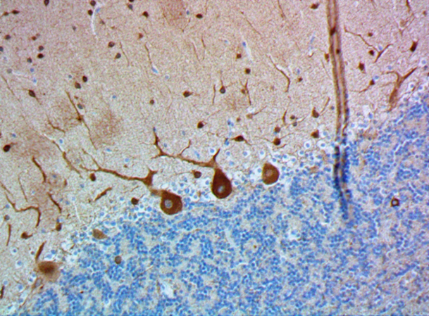
IHC staining of purified anti-Pax6 antibody (clone AD2.35) on formalin-fixed paraffin-embedded mouse brain tissue. Following antigen retrieval using using Sodium Citrate H.I.E.R., the tissue was incubated with 5 µg/mL of the primary antibody overnight at 4°C. BioLegend's Ultra Streptavidin (USA) HRP Detection Kit (Multi-Species, DAB, Cat. No. 929901) was used for detection followed by hematoxylin counterstaining, according to the protocol provided. The image was captured with a 40X objective. Scale bar: 50 µm -

IHC staining of purified anti-Pax6 antibody (clone AD2.35) on frozen mouse cerebellum (A, positive control) and sciatic nerve (B, negative control) tissues. Following fixation (Cat. No. 420801) and permeabilization with 0.5% Triton X-100, the tissue sections were incubated with 5.0 µg/mL of purified anti-Pax6 antibody overnight at 4°C. Alexa Fluor® 594 anti-mouse IgG2a (Cat. No. 407120) antibody was used for 2-hour incubation at room temperature, followed by nuclei counterstain with DAPI (Cat. No. 422801). The slides were then mounted with ProLong™ Gold Antifade Mountant for image acquisition with a 40X objective. Scale bar: 50 μm. -

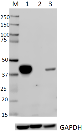
Western blot of purified anti-Pax6 antibody (clone AD2.35). Lane 1: Molecular weight marker; Lane 2: 10 µg of human cerebellum lysate; Lane 3: 20 µg of mouse brain lysate. Lane4: 30 µg of rat brain lysate. The blot was incubated with 5 µg/mL of the primary antibody overnight at 4°C, followed by incubation with HRP-labeled goat anti-mouse IgG (Cat. No. 405306). Enhanced chemiluminescence (Cat. No. 436303) was used as the detection system.
| Cat # | Size | Price | Quantity Check Availability | Save | ||
|---|---|---|---|---|---|---|
| 862001 | 25 µg | 85€ | ||||
| 862002 | 100 µg | 221€ | ||||
Paired box protein Pax6 is a homeobox transcription factor belonging to a family of 9 proteins, which all share a 128 amino-acid DNA binding domain termed “paired domain” (PD). Homologs of Pax proteins exist in worms, flies, frogs, fish and birds. The Pax family has been shown to play important roles in organogenesis and development. The Pax family of proteins modify transcription activity and protein expression levels primarily through binding to the regulatory region of downstream genes. In addition to the PD domain, some Pax proteins, including Pax6, possess another DNA binding domain, a homeodomain (HD), which preferentially binds to sequence with “TAAT” core motif.
Pax6 was first identified by its homology to the Drosophila Paired gene and is mainly expressed in the central nervous system, eye and pancreas. Mutations of Pax6 result in severe eye development abnormalities. Human heterozygous mutations of Pax6 result in aniridia, blindness, cataract and colobomas. In mice, homozygous Pax6 mutations lead to complete loss of ocular structure. In Drosophila, mutation of Pax6 homolog results in an eyeless phenotype. In addition to the eye development, pancreas abnormalities have been also observed in Pax6 deficiency mice.
Product Details
- Verified Reactivity
- Human, Mouse, Rat
- Antibody Type
- Monoclonal
- Host Species
- Mouse
- Immunogen
- KLH conjugated peptide corresponding to amino acid sequence 174-188 of human Pax6.
- Formulation
- Phosphate-buffered solution, pH 7.2, containing 0.09% sodium azide.
- Preparation
- The antibody was purified by affinity chromatography.
- Concentration
- 0.5 mg/ml
- Storage & Handling
- The antibody solution should be stored undiluted between 2°C and 8°C.
- Application
-
ICC - Quality tested
IHC-P, IHC-F, WB - Verified - Recommended Usage
-
Each lot of this antibody is quality control tested by immunocytochemistry. For immunocytochemistry, a concentration range of 2.5 - 5.0 μg/mL is recommended. For formalin-fixed paraffin-embedded immunohistochemistry, a concentration range of 5.0 - 10 µg/ml is suggested. For frozen tissue immunohistochemistry, a concentration range of 1.0 - 10.0 µg/mL is suggested. For Western blotting, the suggested use of this reagent is 5.0 - 10 µg per ml. It is recommended that the reagent be titrated for optimal performance for each application.
- Application Notes
-
Reactivity is weaker in Rat compared to Human and Mouse.
- Product Citations
-
- RRID
-
AB_2801237 (BioLegend Cat. No. 862001)
AB_2801238 (BioLegend Cat. No. 862002)
Antigen Details
- Structure
- Human Pax6 is a 422 amino acid protein with a molecular mass of 46 kD.
- Distribution
-
Tissue distribution: central nervous system, eye, and pancreas
Cellular distribution: nucleus - Function
- Pax6 is involved in the regulation of gene expression.
- Interaction
- Interacts with MAF and MAFB; interacts with TRIM11 Cellular targets: more than 200 target genes, including foxp2, pax6a, gata3, putf3 and more.
- Cell Type
- Neural Stem Cells, Neurons
- Biology Area
- Cell Biology, Neuroscience, Neuroscience Cell Markers, Signal Transduction, Stem Cells, Synaptic Biology, Transcription Factors
- Molecular Family
- Nuclear Markers
- Antigen References
-
- Chi N, et al. 2002. Trends Genet. 18(1): 41-7.
- Coutinho P, et al. 2011. Genome Res. 21(8):1349-59.
- Gene ID
- 5080 View all products for this Gene ID 18508 View all products for this Gene ID 25509 View all products for this Gene ID
- UniProt
- View information about Pax6 on UniProt.org

Related FAQs
Other Formats
View All Pax6 Reagents Request Custom Conjugation| Description | Clone | Applications |
|---|---|---|
| Purified anti-Pax6 | AD2.35 | ICC,IHC-P,IHC-F,WB |
Customers Also Purchased
Compare Data Across All Formats
This data display is provided for general comparisons between formats.
Your actual data may vary due to variations in samples, target cells, instruments and their settings, staining conditions, and other factors.
If you need assistance with selecting the best format contact our expert technical support team.
-
Purified anti-Pax6

Western blot of purified anti-Pax6 antibody (clone AD2.35). ... 
IHC staining of purified anti-Pax6 antibody (clone AD2.35) o... 
IHC staining of purified anti-Pax6 antibody (clone AD2.35) o... 
ICC staining of purified anti-Pax6 antibody (clone AD2.35) o... 
IHC staining of purified anti-Pax6 antibody (clone AD2.35) o...


Follow Us