 Login / Register


- Clone
- AFS98 (See other available formats)
- Regulatory Status
- RUO
- Other Names
- CSF-1R, M-CSFR, c-fms
- Isotype
- Rat IgG2a, κ
- Ave. Rating
- Submit a Review
- Product Citations
- publications

-

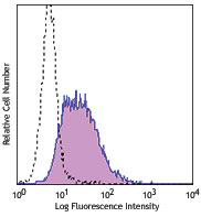
Thioglycolate-elicited BALB/c mouse peritoneal macrophages stained with rat IgG1, k PE and CD11b FITC. -

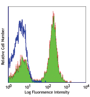
Thioglycolate-elicited BALB/c mouse peritoneal macrophages stained with AFS98 PE and CD11b FITC.
| Cat # | Size | Price | Quantity Check Availability | Save | ||
|---|---|---|---|---|---|---|
| 135501 | 50 µg | 76€ | ||||
| 135502 | 500 µg | 109€ | ||||
CSF-1R, also known as CD115 and M-CSFR, is a single-pass type I membrane protein and member of the platelet-derived growth factor receptor family. This c-fms (Fms proto-oncogene) gene product's natural ligands include M-CSF and IL-34. Structural studies of CD115 have described an Ig-like extracellular domain, a transmembrane domain, an intracellular juxtamembrane domain, a split tyrosine kinase domain, and a C-terminal tail receptor. Receptor activation induces homodimerization in addition to phosphorylation and ubiquitination of intracellular residues. CD115 directly influences tissue macrophage and osteoclast differentiation and proliferation. It is expressed on monocytes/macrophages, peritoneal exudate cells, plasmacytoid and conventional dendritic cells, and osteoclasts.
Product DetailsProduct Details
- Verified Reactivity
- Mouse
- Antibody Type
- Monoclonal
- Host Species
- Rat
- Formulation
- Phosphate-buffered solution, pH 7.2, containing 0.09% sodium azide.
- Preparation
- The antibody was purified by affinity chromatography.
- Concentration
- 0.5 mg/mL
- Storage & Handling
- The antibody solution should be stored undiluted between 2°C and 8°C.
- Application
-
FC - Quality tested
Block - Reported in the literature, not verified in house - Recommended Usage
-
Each lot of this antibody is quality control tested by immunofluorescent staining with flow cytometric analysis. For flow cytometric staining, the suggested use of this reagent is ≤ 0.25 µg per 106 cells in 100 µL volume. It is recommended that the reagent be titrated for optimal performance for each application.
- Application Notes
-
Additional reported applications (for the relevant formats) include: blocking of ligand binding1. The LEAF™ purified antibody (Endotoxin <0.1 EU/µg, Azide-Free, 0.2 µm filtered) is recommended for functional assays.
It has been reported that CD115 can be rapidly internalized, especially when samples are exposed to room temperature. Approximate 33% decrease in CD115 expression has been observed between 0 and 4 hours after sample collection, while overnight incubation of the cells results in complete CD115 downregulation. Pre-treatment with EDTA and low temperatures (2 to 8°C) helps in maintaining surface expression of CD1154. - Application References
- Product Citations
-
- RRID
-
AB_1937292 (BioLegend Cat. No. 135501)
AB_1937293 (BioLegend Cat. No. 135502)
Antigen Details
- Structure
- Ig superfamily, 145 kD
- Distribution
-
Monocytes/macrophages, peritoneal exudate cells, plasmacytoid and conventional dendritic cells, osteoclasts
- Function
- Growth factor receptor, tyrosine kinase
- Ligand/Receptor
- Macrophage colony stimulating factor (M-CSF), IL-34
- Cell Type
- Dendritic cells, Macrophages, Monocytes, Osteoclasts
- Biology Area
- Immunology
- Molecular Family
- CD Molecules, Cytokine/Chemokine Receptors
- Antigen References
-
- Sudo T, et al. 1995 Oncogene 11:2469.
- Murayama T, et al. 1999 Circulation 99:1740.
- Goswami S, et al. 2005 Cancer Res. 65:5278.
- Yu W, et al. 2008 J. Leuko. Biol. 84:852.
- Gene ID
- 12978 View all products for this Gene ID
- UniProt
- View information about CD115 on UniProt.org


Related FAQs
- Why do I have a weak CD115 staining?
-
It has been reported that CD115 can be rapidly internalized, especially when samples are exposed to room temperature. Approximate 33% decrease in CD115 expression has been observed between 0 and 4 hours after sample collection, while overnight incubation of the cells results in complete CD115 downregulation. Pre-treatment with EDTA and low temperatures (2 - 8°C) helps in maintaining surface expression of CD115. In addition, brief fixation of the cells with Fixation Buffer (Cat. No. 420801) for 10 minutes will block CD115 internalization.
Other Formats
View All CD115 Reagents Request Custom ConjugationCustomers Also Purchased
Compare Data Across All Formats
This data display is provided for general comparisons between formats.
Your actual data may vary due to variations in samples, target cells, instruments and their settings, staining conditions, and other factors.
If you need assistance with selecting the best format contact our expert technical support team.
-
Purified anti-mouse CD115 (CSF-1R)

Thioglycolate-elicited BALB/c mouse peritoneal macrophages s... 
Thioglycolate-elicited BALB/c mouse peritoneal macrophages s... -
PE anti-mouse CD115 (CSF-1R)
Thioglycolate-elicited BALB/c mouse peritoneal macrophages s...
Thioglycolate-elicited BALB/c mouse peritoneal macrophages s... -
Biotin anti-mouse CD115 (CSF-1R)

Thioglycolate-elicited BALB/c mouse peritoneal macrophages s... 
-
APC anti-mouse CD115 (CSF-1R)

Thioglycolate-elicted BALB/c mouse peritoneal macrophages st... 
-
Alexa Fluor® 488 anti-mouse CD115 (CSF-1R)

Thioglycolate-elicited C57BL/6 mouse peritoneal macrophages ... -
Brilliant Violet 421™ anti-mouse CD115 (CSF-1R)

Thioglycolate-elicited C57BL/6 mouse peritoneal macrophages ... 
-
Brilliant Violet 605™ anti-mouse CD115 (CSF-1R)

Thioglycolate-elicited C57BL/6 mouse peritoneal macrophages ... 
-
Brilliant Violet 711™ anti-mouse CD115 (CSF-1R)

Thioglycolate-elicited C57BL/6 mouse peritoneal macrophages ... 
-
Alexa Fluor® 594 anti-mouse CD115 (CSF-1R)

C57BL/6 mouse bone marrow derived macrophages were fixed wit... -
Purified anti-mouse CD115 (CSF-1R) (Maxpar® Ready)

Mouse J774A.1 macrophage cells (top) and mouse EL4 T cells (... -
PE/Cyanine7 anti-mouse CD115 (CSF-1R)

Thioglycolate-elicited BALB/c peritoneal macrophages were st... -
PerCP/Cyanine5.5 anti-mouse CD115 (CSF-1R)

Thioglycolate-elicited BALB/c peritoneal macrophages were st... -
PE/Dazzle™ 594 anti-mouse CD115 (CSF-1R)

Thioglycolate-elicited BALB/c mouse peritoneal macrophages w... 
-
Alexa Fluor® 647 anti-mouse CD115 (CSF-1R)

Thioglycolate-elicited BALB/c mouse peritoneal macrophages w... 
-
APC/Cyanine7 anti-mouse CD115 (CSF-1R)

Thioglycolate-elicited BALB/c mouse peritoneal macrophages w... -
TotalSeq™-A0105 anti-mouse CD115 (CSF-1R)
-
APC/Fire™ 750 anti-mouse CD115 (CSF-1R)

Thioglycolate-elicited C57BL/6 mouse peritoneal macrophages ... -
Ultra-LEAF™ Purified anti-mouse CD115 (CSF-1R)
-
TotalSeq™-B0105 anti-mouse CD115 (CSF-1R)
-
TotalSeq™-C0105 anti-mouse CD115 (CSF-1R)
-
Brilliant Violet 785™ anti-mouse CD115 (CSF-1R)

Thioglycolate-elicited BALB/c peritoneal macrophages were st...


Follow Us