 Login / Register


- Clone
- HECA-452 (See other available formats)
- Regulatory Status
- RUO
- Workshop
- V S075
- Other Names
- Cutaneous Lymphocyte-associated Antigen (CLA)
- Isotype
- Rat IgM, κ
- Ave. Rating
- Submit a Review
- Product Citations
- publications

-

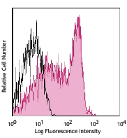
Human peripheral blood lymphocytes were stained with purified CLA (clone HECA-452) (filled histogram) or rat IgM isotype control (open histogram), followed by biotinylated anti-rat IgM and Sav-PE. -

Human paraffin-embedded tonsil tissue slices were prepared with a standard protocol of deparaffinization and rehydration. Antigen retrieval was done with Citrate Buffered 1X (1.0M, pH 7.4) at 95°C for 40 minutes. Tissue was washed with PBS/0.05% Tween 20 twice for five minutes and blocked with 5% FBS and 0.2% gelatin for 30 minutes. Then, the tissue was stained with 10 µg/mL of purified anti-human CLA (clone HECA-452) overnight at 4°C. On the next day, tissue was incubated with Alexa Fluor® 647 goat anti-rat IgM (clone MRM-47) antibody (red) and Alexa Fluor® 594 anti-human CD45RO (Clone UCHL1) antibody (green) for an hour at room temperature. Nuclei were counterstained with DAPI (blue). The image was scanned with a 10X objective and stitched with MetaMorph® software. -

Human paraffin-embedded skin tissue slices were prepared with a standard protocol of deparaffinization and rehydration. Antigen retrieval was done with Citrate Buffered 1X (10mM, pH 6.0) at 95°C for 40 minutes. Tissue was washed with PBS/0.05% Tween 20 twice for five minutes and blocked with 5% FBS and 0.2% gelatin for 30 minutes. Then, the tissue was stained with 10 µg/mL of purified CLA (clone HECA-452) antibody at 4°C overnight. On the next day, tissue was incubated with Alexa Fluor® 647 anti-Rat IgM (clone MRM-47) antibody (red) and Alexa Fluor® 594 anti-human CD44 (clone IM7) antibody (green). The nuclei were conter-stained with DAPI (blue). The image was scanned with a 10X objective and stitched with MetaMorph® software.
| Cat # | Size | Price | Quantity Check Availability | Save | ||
|---|---|---|---|---|---|---|
| 321302 | 100 µg | 96€ | ||||
Cutaneous lymphocyte antigen (CLA) is a 140 kD homodimer protein recognized by a unique mAb, HECA-452. It is expressed on T cells in skin, subsets of peripheral blood memory T cells, NK cells, memory B cells and dendritic cells as well as on monocytes, granulocytes, and activated endothelial cells. CLA is a carbohydrate epitope of sialic acid and fucose-modified P-selectin glycoprotein ligand-1 (PSGL-1), a surface glycoprotein expressed on the majority of peripheral blood leukocytes. CLA is a ligand for E-selectin, P-selectin, and L-selectin. It plays a role in memory lymphocyte homing, tethering, and rolling.
Product DetailsProduct Details
- Verified Reactivity
- Human, Mouse
- Antibody Type
- Monoclonal
- Host Species
- Rat
- Formulation
- Phosphate-buffered solution, pH 7.2, containing 0.09% sodium azide.
- Preparation
- The antibody was purified by affinity chromatography.
- Concentration
- 0.5 mg/ml
- Storage & Handling
- The antibody solution should be stored undiluted between 2°C and 8°C.
- Application
-
FC - Quality tested
IHC-P - Verified
IHC-F - Reported in the literature, not verified in house - Recommended Usage
-
Each lot of this antibody is quality control tested by immunofluorescent staining with flow cytometric analysis. For flow cytometric staining, the suggested use of this reagent is ≤ 2.0 µg per million cells in 100 µl volume. For immunohistochemistry formalin-fixed paraffin-embedded tissue sections, a concentration range of 5.0 - 10.0 µg/ml is suggested. It is recommended that the reagent be titrated for optimal performance for each application.
- Application Notes
-
The HECA-452 antibody cross-reacts with mouse skin homing lymphocytes4. Treatment of activated HUVEC cells with HECA-452 antibody inhibits lymphocyte adhesion. Additional reported applications (for the relevant formats) include: blocking of lymphocyte binding to E-selectin3, and immunohistochemistry1,2 of acetone-fixed frozen sections and formalin-fixed paraffin-embedded sections.
- Application References
-
- Duijvestijn AM, et al. 1988. Am. J. Pathol. 130:147. (IHC)
- Picker LJ, et al. 1991. Nature 349:796. (IHC)
- Berg EL, et al. 1991. J. Exp. Med. 174:1461.
- Borges E, et al. 1997. J. Biol. Chem. 272:28786.
- Ren YL, et al. 2012. Am J Clin Pathol. 138:435. PubMed
- Product Citations
-
- RRID
-
AB_492894 (BioLegend Cat. No. 321302)
Antigen Details
- Structure
- 140 kD, carbohydrate epitope of PSGL-1
- Distribution
-
T cells in skin, subsets of peripheral blood T cells, NK cells, B cells and dendritic cells, monocytes, granulocytes
- Function
- Memory cells tethering and rolling
- Ligand/Receptor
- E-selectin, P-selectin, L-selectin
- Cell Type
- B cells, Dendritic cells, Granulocytes, Monocytes, NK cells, T cells
- Biology Area
- Cell Adhesion, Cell Biology, Immunology
- Molecular Family
- Adhesion Molecules, CD Molecules
- Antigen References
-
1. Picker LJ, et al. 1990. Am. J. Pathol. 136:1053.
2. Berg EL, et al. 1991. J. Exp. Med. 174:1461.
3. Fuhlbrigge RC, et al. 1997. Nature 389:978.
4. Tu L, et al. 1999. J. Exp. Med. 189:241.
5. Yoshino T, et al. 1999. Cell. Immunol. 197:39.
6. Chang SE, et al. 2003. Acta Derm-Venereol. 83:162.
7. Schakel K, et al. 2002. Immunity 17:289.
8. Fuhlbrigge RC, et al. 2002. J. Immunol. 168:5645. - Gene ID
- 6404 View all products for this Gene ID
- UniProt
- View information about CLA on UniProt.org

Related FAQs
Other Formats
View All CLA Reagents Request Custom Conjugation| Description | Clone | Applications |
|---|---|---|
| Purified anti-human/mouse Cutaneous Lymphocyte Antigen (CLA) | HECA-452 | FC,IHC-P,IHC-F |
| FITC anti-human/mouse Cutaneous Lymphocyte Antigen (CLA) | HECA-452 | FC |
| Pacific Blue™ anti-human/mouse Cutaneous Lymphocyte Antigen (CLA) | HECA-452 | FC |
| Alexa Fluor® 647 anti-human/mouse Cutaneous Lymphocyte Antigen (CLA) | HECA-452 | FC |
| PE anti-human/mouse Cutaneous Lymphocyte Antigen (CLA) | HECA-452 | FC |
| PE/Cyanine7 anti-human/mouse Cutaneous Lymphocyte Antigen (CLA) | HECA-452 | FC |
| PerCP/Cyanine5.5 anti-human/mouse Cutaneous Lymphocyte Antigen (CLA) | HECA-452 | FC |
| Alexa Fluor® 594 anti-human/mouse Cutaneous Lymphocyte Antigen (CLA) | HECA-452 | IHC-P |
Customers Also Purchased
Compare Data Across All Formats
This data display is provided for general comparisons between formats.
Your actual data may vary due to variations in samples, target cells, instruments and their settings, staining conditions, and other factors.
If you need assistance with selecting the best format contact our expert technical support team.
-
Purified anti-human/mouse Cutaneous Lymphocyte Antigen (CLA)

Human peripheral blood lymphocytes were stained with purifie... 
Human paraffin-embedded tonsil tissue slices were prepared w... 
Human paraffin-embedded skin tissue slices were prepared wit... -
FITC anti-human/mouse Cutaneous Lymphocyte Antigen (CLA)

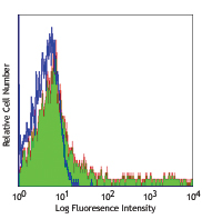
Human peripheral blood lymphocytes stained with HECA-452 FIT... -
Pacific Blue™ anti-human/mouse Cutaneous Lymphocyte Antigen (CLA)

Human peripheral blood lymphocytes stained with HECA-452 Pac... -
Alexa Fluor® 647 anti-human/mouse Cutaneous Lymphocyte Antigen (CLA)

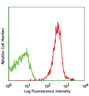
Human peripheral blood lymphocytes were stained with CLA (cl... -
PE anti-human/mouse Cutaneous Lymphocyte Antigen (CLA)

Human peripheral blood lymphocytes were stained with CLA (cl... -
PE/Cyanine7 anti-human/mouse Cutaneous Lymphocyte Antigen (CLA)

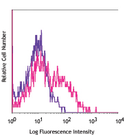
Human peripheral blood lymphocytes were stained with CLA (cl... -
PerCP/Cyanine5.5 anti-human/mouse Cutaneous Lymphocyte Antigen (CLA)

Human peripheral blood granulocytes stained with Cutaneous L... 
Human peripheral blood lymphocytes stained with Cutaneous Ly... -
Alexa Fluor® 594 anti-human/mouse Cutaneous Lymphocyte Antigen (CLA)

Human paraffin-embedded tonsil tissue slices were prepared w... 
Human paraffin-embedded skin tissue slices were prepared wit...


Follow Us