 Login / Register


- Clone
- G10F5 (See other available formats)
- Regulatory Status
- RUO
- Workshop
- VI MA81
- Other Names
- CD67, CGM6, NCA-95, CEACAM8
- Isotype
- Mouse IgM, κ
- Ave. Rating
- Submit a Review
- Product Citations
- publications

-

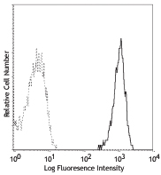
Human peripheral blood granulocytes stained with purified G10F5, followed by anti-mouse Igs FITC -

Human paraffin-embedded spleen tissue slices were prepared with a standard protocol of deparaffinization and rehydration. Antigen retrieval was done with Tris-Buffered Saline 1X with Tween 20 (50 mM, pH 7.6, Cat. No. 925501) at 95°C for 40 minutes. Tissue was washed with PBS/0.05% Tween 20 twice for five minutes and blocked with 5% FBS and 0.2% gelatin for 30 minutes. Then, the tissue was stained with 10 µg/mL of purified anti-human CD66b (clone G10F5) (red) at 4°C overnight. On the next day, tissue was incubated with Alexa Fluor® 647 anti-mouse IgM antibody (clone RMM-1) antibody (red) for an hour. Nuclei were counterstained with DAPI (blue). The image was captured with a 10X objective.
| Cat # | Size | Price | Quantity Check Availability | Save | ||
|---|---|---|---|---|---|---|
| 305102 | 100 µg | 92€ | ||||
CD66b is a 95-100 kD glycosylphosphatidylinositol (GPI)-linked protein also known as CD67, CGM6, and NCA-95. CD66b is a member of the immunoglobulin superfamily, carcinoembryonic antigen (CEA)-like subfamily. CD66b, expressed on granulocytes, has been reported to induce activation in neutrophils and to be involved in heterophilic adhesion with CD66c.
Product DetailsProduct Details
- Verified Reactivity
- Human
- Reported Reactivity
- Chimpanzee
- Antibody Type
- Monoclonal
- Host Species
- Mouse
- Formulation
- Phosphate-buffered solution, pH 7.2, containing 0.09% sodium azide.
- Preparation
- The antibody was purified by affinity chromatography.
- Concentration
- 0.5 mg/ml
- Storage & Handling
- The antibody solution should be stored undiluted between 2°C and 8°C.
- Application
-
FC - Quality tested
IHC-P - Verified - Recommended Usage
-
Each lot of this antibody is quality control tested by immunofluorescent staining with flow cytometric analysis. For flow cytometric staining, the suggested use of this reagent is ≤1.0 µg per million cells in 100 µl volume. For immunohistochemical staining on formalin-fixed paraffin-embedded tissue sections, the suggested use of this reagent is 5.0 - 10 µg per ml. It is recommended that the reagent be titrated for optimal performance for each application.
- Application Notes
-
Additional reported applications (for the relevant formats) include: immunohistochemical staining of acetone-fixed frozen, formalin-fixed paraffin-embedded tissue sections, and spatial biology (IBEX)5,6.
- Application References
-
- Schlossman S, et al. Eds. 1995. Leucocyte Typing V. Oxford University Press. New York.
- Kishimoto T, et al. Eds. 1997. Leucocyte Typing VI. Garland Publishing Inc. London.
- Norling LV, et al. 2012. Arterioscler Thromb Vasc Biol. 32:1970. PubMed
- Meinke P, et al. 2015. Neuroimmunol Discord. 25:127. PubMed
- Radtke AJ, et al. 2020. Proc Natl Acad Sci USA. 117:33455-33465. (SB) PubMed
- Radtke AJ, et al. 2022. Nat Protoc. 17:378-401. (SB) PubMed
- Product Citations
-
- RRID
-
AB_314494 (BioLegend Cat. No. 305102)
Antigen Details
- Structure
- Ig superfamily, CEA antigen group, GPI-linked glycoprotein, 95-100 kD
- Distribution
-
Granulocytes
- Function
- Cell adhesion, neutrophil activation
- Ligand/Receptor
- CD66c
- Cell Type
- Granulocytes, Neutrophils
- Biology Area
- Immunology
- Molecular Family
- Adhesion Molecules, CD Molecules
- Antigen References
-
1. Kuijpers T, et al. 1993. J. Immunol. 151:4934.
2. Kuroki M, et al. 1992. J. Leuk. Biol. 52:551. - Gene ID
- 1088 View all products for this Gene ID
- UniProt
- View information about CD66b on UniProt.org


Other Formats
View All CD66b Reagents Request Custom Conjugation| Description | Clone | Applications |
|---|---|---|
| FITC anti-human CD66b | G10F5 | FC |
| Purified anti-human CD66b | G10F5 | FC,IHC-P |
| Pacific Blue™ anti-human CD66b | G10F5 | FC |
| PE anti-human CD66b | G10F5 | FC |
| PerCP/Cyanine5.5 anti-human CD66b | G10F5 | FC |
| Alexa Fluor® 647 anti-human CD66b | G10F5 | FC,IHC-P,SB |
| Alexa Fluor® 700 anti-human CD66b | G10F5 | FC |
| PE/Cyanine7 anti-human CD66b | G10F5 | FC |
| APC anti-human CD66b | G10F5 | FC |
| Biotin anti-human CD66b | G10F5 | FC |
| PE/Dazzle™ 594 anti-human CD66b | G10F5 | FC |
| Alexa Fluor® 594 anti-human CD66b | G10F5 | IHC-P |
| APC/Cyanine7 anti-human CD66b | G10F5 | FC |
| FITC anti-human CD66b | G10F5 | FC |
| GMP FITC anti-human CD66b | G10F5 | FC |
Customers Also Purchased
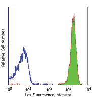
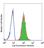
Compare Data Across All Formats
This data display is provided for general comparisons between formats.
Your actual data may vary due to variations in samples, target cells, instruments and their settings, staining conditions, and other factors.
If you need assistance with selecting the best format contact our expert technical support team.
-
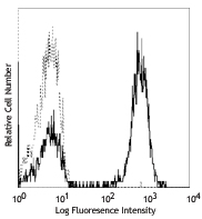
FITC anti-human CD66b

Human peripheral whole blood granulocytes stained with G10F5... -
Purified anti-human CD66b

Human peripheral blood granulocytes stained with purified G1... 
Human paraffin-embedded spleen tissue slices were prepared w... -
Pacific Blue™ anti-human CD66b

Human peripheral blood granulocytes were stained with CD66b ... -
PE anti-human CD66b

Human peripheral blood granulocytes stained with G10F5 PE -
PerCP/Cyanine5.5 anti-human CD66b

Human peripheral blood granulocytes were stained with CD66b ... -
Alexa Fluor® 647 anti-human CD66b

Human peripheral blood granulocytes were stained with CD66b ... 
Human paraffin-embedded spleen tissue was stained with Alexa... 
Confocal image of human lymph node sample acquired using the... -
Alexa Fluor® 700 anti-human CD66b

Human peripheral blood granulocytes were stained with CD66b ... -
PE/Cyanine7 anti-human CD66b

Human peripheral blood granulocytes were stained with CD66b ... -
APC anti-human CD66b

Human peripheral blood granulocytes were stained with CD66b ... -
Biotin anti-human CD66b

Human peripheral blood granulocytes were stained with biotin... -
PE/Dazzle™ 594 anti-human CD66b

Human peripheral blood granulocytes were stained with CD66b ... -
Alexa Fluor® 594 anti-human CD66b

Human paraffin-embedded spleen tissue slices were prepared w... -
APC/Cyanine7 anti-human CD66b

Human peripheral blood granulocytes were stained with CD66b ... -
FITC anti-human CD66b
Typical results from human peripheral blood granulocytes sta... -
GMP FITC anti-human CD66b

Typical results from human peripheral blood granulocytes sta...


Follow Us