 Login / Register


- Clone
- 16B12 (See other available formats)
- Regulatory Status
- RUO
- Other Names
- HA epitope tag, HA1, HA2, hemagglutinin, Hemagglutinin HA1 chain, Hemagglutinin HA2 chain, YPYDVPDYA, Hemagglutinin tag
- Isotype
- Mouse IgG1, κ
- Ave. Rating
- Submit a Review
- Product Citations
- publications

-

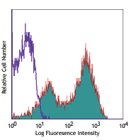
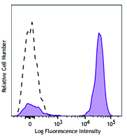
Transfected RBL1 cells expressing the HA tag on the cell surface were stained with anti-HA.11 Epitope Tag (clone 16B12) PE/Dazzle™ 594 (filled histogram) or mouse IgG1, κ PE/Dazzle™ 594 isotype control (open histogram). -

CHO-K1 cells (open histogram) or HA tag stably transfected cells (filled histogram) were fixed with Fixation Buffer (Cat. No. 420801), permeabilized with True-Phos™ Perm Buffer (Cat. No. 425401), then intracellularly stained with HA.11 Epitope Tag (16B12) PE/Dazzle™ 594.
| Cat # | Size | Price | Quantity Check Availability | Save | ||
|---|---|---|---|---|---|---|
| 901529 | 25 µg | 151€ | ||||
| 901530 | 100 µg | 372€ | ||||
The HA tag (hemagglutinin) is an amino acid sequence derived from the human influenza hemagglutinin surface glycoprotein, corresponding to amino acids 98-106. It is commonly used as a tag to facilitate detection, isolation, and purification of proteins. The full amino acid sequence is: YPYDVPDYA.
Product DetailsProduct Details
- Antibody Type
- Monoclonal
- Host Species
- Mouse
- Immunogen
- Monoclonal antibody HA.11 was raised against the twelve amino acid peptide CYPYDVPDYASL.
- Formulation
- Phosphate-buffered solution, pH 7.2, containing 0.09% sodium azide.
- Preparation
- The antibody was purified by affinity chromatography and conjugated with PE/Dazzle™ 594 under optimal conditions.
- Concentration
- 0.2 mg/ml
- Storage & Handling
- The antibody solution should be stored undiluted between 2°C and 8°C, and protected from prolonged exposure to light. Do not freeze.
- Application
-
FC - Quality tested
ICFC - Verified - Recommended Usage
-
Each lot of this antibody is quality control tested by immunofluorescent staining with flow cytometric analysis. For flow cytometric staining, the suggested use of this reagent is ≤ 0.25 µg per million cells in 100 µL volume. For intracellular flow cytometric staining using our True-Phos™ Perm Buffer in Cell Suspensions Protocol, the suggested use of this reagent is ≤ 0.125 µg per million cells in 100 µL volume. It is recommended that the reagent be titrated for optimal performance for each application.
* PE/Dazzle™ 594 has a maximum excitation of 566 nm and a maximum emission of 610 nm. - Excitation Laser
-
Blue Laser (488 nm)
Green Laser (532 nm)/Yellow-Green Laser (561 nm)
- Application Notes
-
Additional tested and reported applications of the 16B12 clone for the relevant formats include: western blot (WB), immunocytochemistry (ICC), immunoprecipitation (IP), and flow cytometry (FC).
*Our Posi-Tag Control Protein (931301) can be used as a helpful positive control for this antibody.
This second-generation HA antibody is an excellent substitute for the 12CA5 monoclonal antibody. The HA.11 antibody recognizes the influenza hemagglutinin epitope (YPYDVPDYA) which has been used extensively as a general epitope tag in expression vectors. The extreme specificity of the antibody allows unambiguous identification and quantitative analysis of the tagged protein. The HA.11 antibody recognizes HA epitopes located in the middle of protein sequences as well as at the N- or C-terminus. - Application References
-
- Kim JY, et al. 2003. J Neurosci. 23:5561. (IP, WB)
- Helliwell SB, et al. 2001. J Cell Biol. 153:649. (WB)
- Bennett BD, et al. 2000. J Biol Chem. 275:37712. (IF, IP, WB)
- Royer Y, et al. 2005. J. Biol. Chem. 29:27251. (FC)
- Smith BA, et al. 2012. Genes Cancer. 3:550. (IHC) PubMed
- Hogarth C, et al. 2015. Biol Reprod. 93:19. PubMed
- Görtz D, et al. 2015. Sci Rep. 5:14685. PubMed
- Wilson C, et al. 2015. PLoS One. 10:0139579. PubMed
- Smith B, et al. 2012. Genes Cancer. 3:550-563. PubMed
- Liu Z, et al. 2016. Nature. 530:98-102. PubMed
- Thoms M, et al. 2016. Nucleic Acids Res. 44:926-39. PubMed
- Kim Y, et al. 2016. Nat Commun. 7:10347. PubMed
- Rodríguez-Escudero M, et al. 2016. PLoS One. 11:0148032. PubMed
- Lehmann W, et al. 2016. Nat Commun. 7:10498. PubMed
- Testoni E, et al. 2016. EMBO Mol Med. 8: 105-16. PubMed
- Padilla S, et al. 2016. Nat Neurosci. 10.1038/nn.4274. PubMed
- Martins J, et al. 2016. J Cell Sci. 129:1271-82. PubMed
- Matak P, et al. 2016. Proc Natl Acad Sci U S A. 113:3428-35. PubMed
- Starokadomskyy P, et al. 2016. Nat Immunol. 17:495-504. PubMed
- Mitxelena J, et al. 2016. Nucleic Acids Res. 44:5557-70. PubMed
- Thongthip S, et al. 2016. Genes Dev. 30:645-59. PubMed
- Aaes T, et al. 2016. Cell Rep. 15:274-87. PubMed
- Hodge C, et al. 2016. J Biol Chem. 291:9396-9410. PubMed
- Alagramam K, et al. 2016. Nat Chem Biol. 12:444-51. PubMed
- Veit G, et al. 2016. PLoS Biol. 14:1002462. PubMed
- Lee B, et al. 2016. Development. 143:1721-31. PubMed
- Douchi D, et al. 2016. Plant Cell. 28:1182-99. PubMed
- Avgousti D, et al. 2016. Nature. 535:173-77. PubMed
- Shin H, et al. 2015. Nature. 534:553-7. PubMed
- Gross G, et al. 2016. Nat Methods. 10.1038/nmeth.3894. PubMed
- Dick M, et al. 2016. Nat Commun. 7:11929. PubMed
- Aldrin-Kirk P, et al. 2016. Neuron. 90:955-68. PubMed
- Fan R, et al. 2016. Nat Med. 22:780-91. PubMed
- Rowald K, et al. 2016. Cell Rep. 15:2679-91. PubMed
- Rampal R, Awasthi A, Ahuja V 2016. Development. 143:2334-43. PubMed
- Faden F, et al. 2016. Nat Commun. 7:12202. PubMed
- Lawson C, et al. 2016. Cancer Res. 76: 3826-37. PubMed
- Szargel R, et al. 2016. Hum Mol Genet. 10.1093/hmg/ddw189.. PubMed
- Damez-Werno D, et al. 2016. Proc Natl Acad Sci U S A. 113: 9623-28. PubMed
- Su P, et al. 2016. J Immunol. 197: 1054-64. PubMed
- Morozumi Y, et al. 2016. J Mol Cell Biol. 8:349-62. PubMed
- Miura Y, et al. 2016. Biochem J. 473:2591-602. PubMed
- Pashkova N, et al. 2016. Cell Rep. 17:303-15. PubMed
- Sanchez M, et al. 2016. J Biol Chem. 291:19760-73. PubMed
- Hwang J, Lee J, Pallas D 2016. J Biol Chem. 291:21008-19. PubMed
- Jiménez-Canino R, et al. 2016. J Biol Chem. 291:19068-78. PubMed
- Barquilla A, et al. 2016. Mol Biol Cell. 27:2757-70. PubMed
- Yadav S, et al. 2016. Sci Rep. 6:34100. PubMed
- Hashimoto A, et al. 2016. Oncogenesis. 0.388194444. PubMed
- Guirouilh-Barbat J, et al. 2016. PLoS Genet. 12:e1006230. PubMed
- Gómez-H L, et al. 2016. Nat Commun. 7:13298. PubMed
- Rademacher N, et al. 2016. Sci Rep. 6:35283. PubMed
- Lin Z, et al. 2016. Nat Genet. 10.1038/ng.3701. PubMed
- Kaczmarska Z, et al. 2016. Nat Chem Biol. 10.1038/nchembio.2217. PubMed
- Zee B, et al. 2016. PLoS One. 11:e0163820. PubMed
- Despras E, et al. 2016. Nat Commun. 7:13326. PubMed
- Zhang H, et al. 2016. J Neurosci. 36:11837-50. PubMed
- Zhang M, et al. 2016. Cell Res. 26:1302-19. PubMed
- Baehr C, et al. 2016. J Biol Chem. 291:26875-85. PubMed
- Boehm E, et al. 2016. J Biol Chem. 291:25877-87. PubMed
- Gail Kilroy, et al. 2016. J Biol Chem. 291:27289-97. PubMed
- Hongchen Cai, Aimin Liu 2016. Proc Natl Acad Sci U S A. 113:14751-6. PubMed
- Tam K, et al. 2016. MBio. 7:e02024-16. PubMed
- Hanke L, et al. 2016. MBio. 7(6). pii: e01569-16. PubMed
- Stoeber M, et al. 2016. Proc Natl Acad Sci U S A. 113(50):E8069-E8078. PubMed
- Gu Q, et al. 2016. J Virol. 90:10545-57. PubMed
- Ramachandran S, et al. 2016. J Biol Chem. 291: 25489-504. PubMed
- Kaufmann T, et al. 2016. J Cell Sci. 129:4607-21. PubMed
- Piwko W, et al. 2016. EMBO J. 35:2584-601. PubMed
- Matsuo Y, et al. 2016. J Cell Sci. 129:4592-606. PubMed
- Athmer J, et al. 2017. MBio. 10.1128/mBio.02320-16. PubMed
- Gao L, et al. 2017. J Cell Sci. 130:396-405. PubMed
- Gong Y, et al. 2017. Genes Dev. 31:46-58. PubMed
- K Kataoka, K Mochizuki 2017. J Cell Sci. 130:480-9. PubMed
- Liszczak G, et al. 2017. Proc Natl Acad Sci U S A. 114:681-6. PubMed
- Haggie P, et al. 2017. J Biol Chem. 292:771-85. PubMed
- T Taetzsch, et al. 2017. J Neurosci. 37:70-82. PubMed
- Yagita Y, et al. 2017. J Biol Chem. 292:691-705. PubMed
- Chong Jiang, et al. 2017. J Biol Chem. 292:3137-45. PubMed
- Maeda R, et al. 2017. J Biol Chem. 292:3201-12. PubMed
- Davis RB, et al. 2017. JCI Insight. 2:e92465. PubMed
- Daniel JA, et al. 2017. Elife. 6:e26338. PubMed
- Oishi A, et al. 2017. Sci Rep. 7:8990. PubMed
- RRID
-
AB_2734663 (BioLegend Cat. No. 901529)
AB_2734664 (BioLegend Cat. No. 901530)
Antigen Details
- Biology Area
- Cell Biology
- Gene ID
- NA
- UniProt
- View information about HA.11 on UniProt.org


Related FAQs
Other Formats
View All HA.11 Reagents Request Custom ConjugationCustomers Also Purchased
Compare Data Across All Formats
This data display is provided for general comparisons between formats.
Your actual data may vary due to variations in samples, target cells, instruments and their settings, staining conditions, and other factors.
If you need assistance with selecting the best format contact our expert technical support team.
-
Anti-HA.11 Epitope Tag Affinity Matrix
-
Alexa Fluor® 488 anti-HA.11 Epitope Tag

HA tagged CHO cells stained with Alexa Fluor® 488 Labeled An... 
Transfected RBL1 cells expressing the HA tag on the cell sur... -
Alexa Fluor® 594 anti-HA.11 Epitope Tag

Staining of HA.11 Clone 16B12 Monoclonal Antibody, Alexa Flu... -
Anti-HA.11 Epitope Tag

Immunofluorescence of HA.11 tagged Sbhlp protein. Photo cour... 
Western blot analysis of cell lysates from CHO and CHO HA st... 
Immunoprecipitation of HA protein from CHO and CHO-HA stable... 
Immunofluorescence staining of HeLa cells transfected withou... -
Biotin anti-HA.11 Epitope Tag

Total lysates (15 µg protein) from CHO (lane 1) and CHO HA s... 
Immunofluorescence of HA-tagged plasmid-transfected A549 cel... -
FITC anti-HA.11 Epitope Tag

Transfected RBL1 cells expressing the HA tag on the cell sur... -
Purified anti-HA.11 Epitope Tag

Western blot of Clone 16B12. Lane 1: Molecular weight marker... 
Staining of Clone 16B12 on methanol fixed CHO cells transfec... 
Immunoprecipitation of HA protein from CHO and CHO-HA stable... 
Transfected RBL1 cells expressing the HA tag on the cell sur... -
Alexa Fluor® 647 anti-HA.11 Epitope Tag

HA tag stably transfected CHO cells were fixed with ice cold... 
Transfected RBL1 cells expressing the HA tag on the cell sur... -
PE anti-HA.11 Epitope Tag

CHO-K1 cells (open histogram) or HA tag stably transfected c... 
Transfected RBL1 cells expressing the HA tag on the cell sur... -
Direct-Blot™ HRP anti-HA.11 Epitope Tag

Total cell lysate from CHO (lane 1) and CHO stably transfect... -
Ultra-LEAF™ Purified anti-HA.11 Epitope Tag

Total cell lysate (15 µg protein) from CHO (lane 1) and CHO ... 
Transfected RBL1 cells expressing the HA tag on the cell sur... -
Brilliant Violet 421™ anti-HA.11 Epitope Tag

CHO cells stably transfected with HA tag were fixed with 4% ... 
Transfected RBL1 cells expressing the HA tag on the cell sur... -
PE/Dazzle™ 594 anti-HA.11 Epitope Tag

CHO-K1 cells (open histogram) or HA tag stably transfected c... 
Transfected RBL1 cells expressing the HA tag on the cell sur... -
PE/Cyanine7 anti-HA.11 Epitope Tag

CHO-K1 cells (open histogram) or HA tag stably transfected c... 
Transfected RBL1 cells expressing the HA tag on the cell sur... -
Pacific Blue™ anti-HA.11 Epitope Tag

CHO-K1 cells (open histogram) or HA tag stably transfected c... 
Transfected RBL1 cells expressing the HA tag on the cell sur... -
APC anti-HA.11 Epitope Tag

CHO-K1 cells (open histogram) or HA tag stably transfected c... 
Transfected RBL1 cells expressing the HA tag on the cell sur... -
PerCP/Cyanine5.5 anti-HA.11 Epitope Tag

CHO-K1 cells (open histogram) or HA tag stably transfected c... 
Transfected RBL1 cells expressing the HA tag on the cell sur... -
TotalSeq™-C1131 anti-HA.11 Epitope Tag
-
TotalSeq™-A1131 anti-HA.11 Epitope Tag
-
TotalSeq™-B1131 anti-HA.11 Epitope Tag


Follow Us