 Login / Register


- Clone
- BVD3-1F9 (See other available formats)
- Regulatory Status
- RUO
- Other Names
- Interleukin-3, Burst promoting activity, Eosinophil colony stimulating factor (Eo-CSF), Hematopoietic cell growth factor (HCGF), Mast (MGF/MCGF), Multi-colony stimulating (Multi-CSF), P cell stimulating activity (PCSA), Thy1 inducing factor
- Isotype
- Rat IgG1, κ
- Ave. Rating
- Submit a Review
- Product Citations
- publications


| Cat # | Size | Price | Quantity Check Availability | Save | ||
|---|---|---|---|---|---|---|
| 500604 | 500 µg | 263€ | ||||
IL-3 is a highly species-specific pleiotropic factor produced primarily by activated T cells though also by mast cells, keratinocytes, and astrocytes, which stimulates colony formation of megakaryocytes, neutrophils, and macrophages from bone marrow cultures. The BVD3-1F9 antibody can neutralize the bioactivity of natural or recombinant IL-3.
Product DetailsProduct Details
- Verified Reactivity
- Human
- Antibody Type
- Monoclonal
- Host Species
- Rat
- Immunogen
- Yeast-expressed, recombinant human IL-3
- Formulation
- Phosphate-buffered solution, pH 7.2, containing 0.09% sodium azide.
- Preparation
- The antibody was purified by affinity chromatography, and conjugated with biotin under optimal conditions.
- Concentration
- 0.5 mg/ml
- Storage & Handling
- The antibody solution should be stored undiluted between 2°C and 8°C. Do not freeze.
- Application
-
ELISA Detection, ELISPOT Detection
- Recommended Usage
-
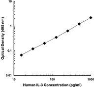
Each lot of this antibody is quality control tested by ELISA assay. For use as an ELISA detection antibody, a concentration range of 0.5-2.0 µg/ml is recommended. To obtain a linear standard curve, serial dilutions of human IL-3 protein ranging from 2000 to 15 pg/ml are recommended for each ELISA plate. It is recommended that the reagent be titrated for optimal performance for each application.
- Application Notes
-
ELISA1-3,6 or ELISPOT4 Detection: The biotinylated BVD3-1F9 antibody is useful as a detection antibody for a sandwich ELISA or ELISPOT assay, when used in conjunction with purified BVD8-3G11 (Cat. No. 500502) antibody as the capture antibody.
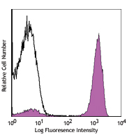
Flow Cytometry: The fluorochrome-labelled BVD3-1F9 antibody is useful for intracellular immunofluorescent staining and flow cytometric analysis to identify IL-3 -producing cells within mixed cell populations. For intracellular cytokine staining protocol, please visit www.biolegend.com and click on the support section.
Additional reported applications (for the relevant formats) include: immunoprecipitation6, Western blotting6, neutralization1,6, immunohistochemical staining5,7 of paraformaldehyde-fixed, saponin-treated frozen tissue sections, and immunocytochemistry. - Application References
-
- Abrams J, et al. 1992. Immunological Reviews 127:5.
- Abrams J, et al. 1994. Eosinophils in Allergy and Inflammation. Marcel Dekker New York. p.133.
- Abrams J. 1995. Curr. Prot. Immunol.. 6.20.
- Mahanty S, et al. 1992. J. Immunol. 148:3567.
- Andersson U, et al. 1993. Detection and quantification of gene expression. New York: Springer-Verlag.
- Kaushansky K. 1992. J. Clin. Invest. 90:1879.
- Andersson J, et al. 1994. Immunology 83:16.
- RRID
-
AB_2123713 (BioLegend Cat. No. 500604)
Antigen Details
- Structure
- Cytokine; 14-30 kD (Mammalian)
- Bioactivity
- Proliferation/differentiation of almost all types of hematopoietic progenitors into granulocytes, macrophages, erythroid cells, megakaryocytes, mast cell colonies; induces expression of 20-α-steroid dehydrogenase, histidine, ornithine decarboxylase
- Cell Sources
- Activated T cells, mast cells, eosinophils, keratinocytes, NK cells, endothelial cells
- Cell Targets
- Erythroid cells, megakaryocytes, neutrophils, eosinophils, basophils, mast cells, monocytic lineages
- Receptors
- IL-3R, α subunit (CD123), common β subunit (CDw131)
- Biology Area
- Cell Biology, Stem Cells
- Molecular Family
- Cytokines/Chemokines, Growth Factors
- Antigen References
-
1. Fitzgerald K, et al. Eds. 2001. The Cytokine FactsBook. Academic Press San Diego.
2. Frendl G. 1992. Int. J. Immunopharmacol. 14:421.
3. Ihle J. 1992. Chem. Immunology 51:65. - Regulation
- Upregulated by T cell stimulation; downregulated by glucocorticoids
- Gene ID
- 3562 View all products for this Gene ID
- UniProt
- View information about IL-3 on UniProt.org
Related Pages & Pathways
Pathways


Related FAQs
- How many biotin molecules are per antibody structure?
- We don't routinely measure the number of biotins with our antibody products but the number of biotin molecules range from 3-6 molecules per antibody.
Other Formats
View All IL-3 Reagents Request Custom Conjugation| Description | Clone | Applications |
|---|---|---|
| Biotin anti-human IL-3 | BVD3-1F9 | ELISA Detection,ELISPOT Detection |
| PE anti-human IL-3 | BVD3-1F9 | ICFC |
Customers Also Purchased
Compare Data Across All Formats
This data display is provided for general comparisons between formats.
Your actual data may vary due to variations in samples, target cells, instruments and their settings, staining conditions, and other factors.
If you need assistance with selecting the best format contact our expert technical support team.
-
Biotin anti-human IL-3

-
PE anti-human IL-3

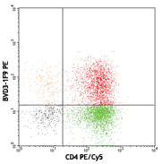
Enriched human CD4+ T cells were stimulated with PMA+ionomyc...


Follow Us