 Login / Register


- Clone
- DX2 (See other available formats)
- Regulatory Status
- RUO
- Workshop
- VI C-64
- Other Names
- Fas, APO-1, TNFRSF6
- Isotype
- Mouse IgG1, κ
- Ave. Rating
- Submit a Review
- Product Citations
- publications

-

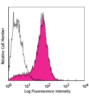
Human peripheral blood lymphocytes were stained with CD95 (Fas) (clone DX2) APC/Cyanine7 (filled histogram) or Mouse IgG1, ? APC/Cyanine7 isotype control (open histogram).
| Cat # | Size | Price | Quantity Check Availability | Save | ||
|---|---|---|---|---|---|---|
| 305635 | 25 tests | 118€ | ||||
| 305636 | 100 tests | 259€ | ||||
CD95 is a 45 kD single chain type I glycoprotein also known as Fas, APO-1, and TNFRSF6. It is a member of the TNF receptor superfamily. CD95 is expressed on T and B lymphocytes, monocytes, neutrophils, and fibroblasts. CD95 expression is upregulated by activation. The extracellular region of CD95 binds to CD178 (Fas ligand). CD178 binding to CD95 induces apoptosis and has been shown to play a role in the maintenance of peripheral tolerance.
Product DetailsProduct Details
- Verified Reactivity
- Human, Cynomolgus, Rhesus
- Reported Reactivity
- African Green, Baboon, Capuchin Monkey, Chimpanzee, Common Marmoset, Cotton-topped Tamarin, Pigtailed Macaque, Sooty Mangabey
- Antibody Type
- Monoclonal
- Host Species
- Mouse
- Immunogen
- CD95 transfected L cells
- Formulation
- Phosphate-buffered solution, pH 7.2, containing 0.09% sodium azide and BSA (origin USA)
- Preparation
- The antibody was purified by affinity chromatography and conjugated with APC/Cyanine7 under optimal conditions.
- Concentration
- Lot-specific (to obtain lot-specific concentration and expiration, please enter the lot number in our Certificate of Analysis online tool.)
- Storage & Handling
- The antibody solution should be stored undiluted between 2°C and 8°C, and protected from prolonged exposure to light. Do not freeze.
- Application
-
FC - Quality tested
- Recommended Usage
-
Each lot of this antibody is quality control tested by immunofluorescent staining with flow cytometric analysis. For flow cytometric staining, the suggested use of this reagent is 5 µl per million cells in 100 µl staining volume or 5 µl per 100 µl of whole blood.
- Excitation Laser
-
Red Laser (633 nm)
- Application Notes
-
The DX2 antibody is useful for inducing apoptosis of Fas-positive cells. Additional reported applications (for the relevant formats) include: in vitro induction of apoptosis3 (DX2 antibody is required to be cross-linked for effective induction of apoptosis) and immunohistochemical staining4,5 of acetone-fixed frozen tissue sections and formalin-fixed paraffin-embedded tissue sections. The Ultra-LEAF™ purified antibody (Endotoxin < 0.01 EU/µg, Azide-Free, 0.2 µm filtered) is recommended for functional assays (Cat. No. 305655 and 305656).
Note: EOS9.1 antibody can induce apoptosis without cross-linking. - Additional Product Notes
- BioLegend is in the process of converting the name APC/Cy7 to APC/Cyanine7. The dye molecule remains the same, so you should expect the same quality and performance from our APC/Cyanine7 products. Please contact Technical Service if you have any questions.
- Application References
-
- Schlossman S, et al. Eds.1995. Leucocyte Typing V. Oxford University Press. New York.
- Kishimoto T, et al. Eds. 1997. Leucocyte Typing VI. Garland Publishing Inc. New York.
- Cifone M, et al. 1994. J. Exp. Med. 180:1547. (Apop)
- Zietz C, et al. 2001. Am. J. Pathol. 159:963. (IHC)
- Sergi C, et al. 2000. Am. J. Pathol. 156:1589. (IHC)
- Xie S, et al. 2010. J. Immunol. 184:2289. (FC) PubMed
- Yoshino N, et al. 2000. Exp. Anim. (Tokyo) 49:97. (FC)
- Sestak K, et al. 2007. Vet. Immunol. Immunopathol. 119:21.
- Rout N, et al. 2010. PLoS One 5:e9787. (FC)
- Dixit N, et al. 2012. J. Immunol. 189:5954. PubMed
- Product Citations
-
- RRID
-
AB_2566108 (BioLegend Cat. No. 305635)
AB_2566111 (BioLegend Cat. No. 305636)
Antigen Details
- Structure
- TNFR superfamily, type I transmembrane protein, 45 kD
- Distribution
-
T cells and B cells, monocytes, neutrophils, fibroblasts, expression level upregulated on activated lymphocytes
- Function
- Mediates apoptosis
- Ligand/Receptor
- Fas ligand (CD178)
- Cell Type
- B cells, Fibroblasts, Lymphocytes, Monocytes, Neutrophils, T cells
- Biology Area
- Apoptosis/Tumor Suppressors/Cell Death, Cell Biology, Immunology, Neuroscience
- Molecular Family
- CD Molecules
- Antigen References
-
1. Krammer P, et al. 1994. Immunol. Rev. 142:175.
2. Nagata S, et al. 1995. Science 267:1449. - Gene ID
- 355 View all products for this Gene ID
- UniProt
- View information about CD95 on UniProt.org
Related Pages & Pathways
Pathways


Related FAQs
Other Formats
View All CD95 Reagents Request Custom ConjugationCustomers Also Purchased
Compare Data Across All Formats
This data display is provided for general comparisons between formats.
Your actual data may vary due to variations in samples, target cells, instruments and their settings, staining conditions, and other factors.
If you need assistance with selecting the best format contact our expert technical support team.
-
APC anti-human CD95 (Fas)

Human peripheral blood lymphocytes stained with DX2 APC -
Biotin anti-human CD95 (Fas)

Human peripheral blood lymphocytes stained with biotinylated... -
FITC anti-human CD95 (Fas)

Human peripheral blood lymphocytes stained with DX2 FITC -
PE anti-human CD95 (Fas)

Human peripheral blood lymphocytes stained with DX2 PE -
PE/Cyanine5 anti-human CD95 (Fas)

Human peripheral blood lymphocytes stained with DX2 PE/Cyani... -
Purified anti-human CD95 (Fas)

Human peripheral blood lymphocytes stainedwith DX2 PE/Cy5 
MCF7 breast cancer cells were stained with anti-human CD95 (... -
Alexa Fluor® 488 anti-human CD95 (Fas)

Human peripheral blood lymphocytes stained with DX2 Alexa Fl... -
Alexa Fluor® 647 anti-human CD95 (Fas)

Human peripheral blood lymphocytes stained with DX2 Alexa Fl... -
Brilliant Violet 421™ anti-human CD95 (Fas)

Human peripheral blood lymphocytes were stained with CD95 (c... -
Pacific Blue™ anti-human CD95 (Fas)

Human peripheral blood lymphocytes stained with DX2 Pacific ... -
PE/Cyanine7 anti-human CD95 (Fas)

Human peripheral blood lymphocytes stained with DX2 PE/Cyani... -
Brilliant Violet 605™ anti-human CD95 (Fas)

Human peripheral blood lymphocytes were stained with CD95 (c... -
PerCP/Cyanine5.5 anti-human CD95 (Fas)

Human peripheral blood lymphocytes were stained with CD95 (c... -
Purified anti-human CD95 (Fas) (Maxpar® Ready)

Human PBMCs stained with 154Sm-anti-CD45 (HI30) and 164Dγ-an... -
PE/Dazzle™ 594 anti-human CD95 (Fas)

Human peripheral blood lymphocytes were stained with CD95 (c... -
APC/Fire™ 750 anti-human CD95 (Fas)

Human peripheral blood lymphocytes were stained with CD95 (c... -
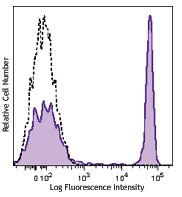
APC/Cyanine7 anti-human CD95 (Fas)

Human peripheral blood lymphocytes were stained with CD95 (F... -
Brilliant Violet 510™ anti-human CD95 (Fas)

Human peripheral blood lymphocytes were stained with CD95 (c... -
Brilliant Violet 711™ anti-human CD95 (Fas)

Human peripheral blood lymphocytes were stained with CD95 (c... -
Brilliant Violet 785™ anti-human CD95 (Fas)

Human peripheral blood lymphocytes were stained with CD95 (c... -
Brilliant Violet 650™ anti-human CD95 (Fas)

Human peripheral blood lymphocytes were stained with CD95 (c... -
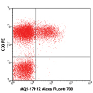
Alexa Fluor® 700 anti-human CD95 (Fas)

Human peripheral blood lymphocytes were stained with CD95 (F... -
TotalSeq™-A0156 anti-human CD95 (Fas)
-
TotalSeq™-C0156 anti-human CD95 (Fas)
-
TotalSeq™-B0156 anti-human CD95 (Fas)
-
Ultra-LEAF™ Purified anti-human CD95 (Fas)

Human peripheral blood lymphocytes stained with Ultra-LEAF™ ...
MCF7 breast cancer cells were stained with anti-human CD95 (... -
TotalSeq™-D0156 anti-human CD95 (Fas)
-
PE/Fire™ 640 anti-human CD95 (Fas)

Human peripheral blood lymphocytes were stained with anti-hu... -
KIRAVIA Blue 520™ anti-human CD95 (Fas)

Human peripheral blood lymphocytes were stained with anti-hu... -
APC/Fire™ 810 anti-human CD95 (Fas) Antibody

Human peripheral lymphocytes were stained with anti-human CD... -
PE anti-human CD95

Typical results from human peripheral blood lymphocytes stai... -
Spark YG™ 593 anti-human CD95 (Fas)

Human peripheral blood monocytes were stained with anti-huma... 
Human peripheral blood monocytes were stained with anti-huma... -
PE/Fire™ 700 anti-human CD95 (Fas)

Human peripheral blood monocytes were stained with anti-huma... 
Human peripheral blood monocytes were stained with anti-huma... -
PerCP/Fire™ 780 anti-human CD95 (Fas)

Human peripheral blood monocytes were stained with anti-huma... 
Human peripheral blood monocytes were stained with anti-huma... -
Spark Blue™ 574 anti-human CD95 (Fas)

Human peripheral blood lymphocytes were stained with anti-hu...


Follow Us