 Login / Register


- Clone
- P82H9 (See other available formats)
- Regulatory Status
- RUO
- Other Names
- MBP, Golli-MBP, myelin A1 protein, myelin membrane encephalitogenic protein
- Isotype
- Mouse IgG1, κ
- Ave. Rating
- Submit a Review
- Product Citations
- publications

-

IHC staining of Alexa Fluor® 488 anti-Myelin Basic Protein antibody (clone P82H9) on formalin-fixed paraffin-embedded rat brain tissue. Following antigen retrieval using Sodium Citrate H.I.E.R. (Cat. No.928602), the tissue was incubated with 5 µg/mL of the primary antibody overnight at 4°C. Nuclei were counterstained with Hoechst, and the slide was mounted with ProLong™ Gold Antifade Mountant. The image was captured with a 40X objective. Scale Bar: 50 µm -

IHC staining of Alexa Fluor® 488 anti-Myelin Basic Protein antibody (clone P82H9) on formalin-fixed paraffin-embedded mouse brain tissue. Following antigen retrieval using Sodium Citrate H.I.E.R. (Cat. No.928602), the tissue was incubated with 5 µg/mL of the primary antibody overnight at 4°C. Nuclei were counterstained with Hoechst, and the slide was mounted with ProLong™ Gold Antifade Mountant. The image was captured with a 40X objective. Scale Bar: 50 µm -

IHC staining of Alexa Fluor® 488 anti-Myelin Basic Protein antibody (clone P82H9) on formalin-fixed paraffin-embedded human brain tissue. Following antigen retrieval using Sodium Citrate H.I.E.R. (Cat. No.928602), the tissue was incubated with 10 µg/mL of the primary antibody overnight at 4°C. Nuclei were counterstained with Hoechst, and the slide was mounted with ProLong™ Gold Antifade Mountant. The image was captured with a 40X objective. Scale Bar: 50 µm -
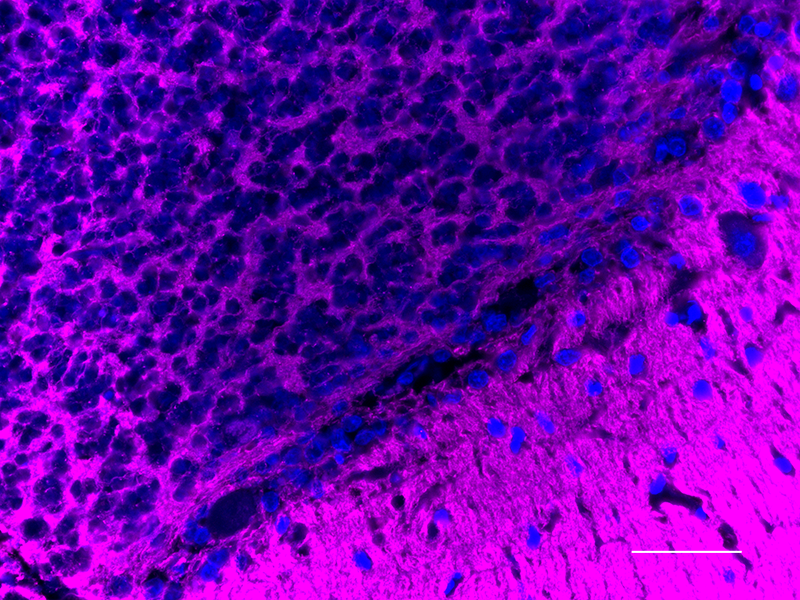
Formalin-fixed, 300 micron-thick mouse brain (dentate gyrus) section was blocked, permeabilized and stained overnight with Myelin Basic Protein (clone P82H9) Alexa Fluor® 488 (green) and MAP2 (clone SMI 52) Alexa Fluor® 647 (magenta) both at 5 µg/mL, optically cleared, and analyzed on a confocal microscope.

| Cat # | Size | Price | Quantity Check Availability | Save | ||
|---|---|---|---|---|---|---|
| 850907 | 25 µg | 128€ | ||||
| 850908 | 100 µg | 259€ | ||||
Myelin basic protein (MBP) is a protein involved in the myelination of nerves in the central nervous system (CNS). MBP functions to maintain the correct structure of myelin, through its interaction with the lipids in the myelin membrane. The myelin sheath is made up of MBP and lipids, and acts as an insulator to increase the velocity of axonal impulse conduction. MBP plays a central role in the demyelinating disease multiple sclerosis (MS). A hallmark of the disease is the loss of the myelin sheath surrounding nerves, thought to be induced by antibodies against MBP.
Product DetailsProduct Details
- Verified Reactivity
- Human, Mouse, Rat
- Antibody Type
- Monoclonal
- Host Species
- Mouse
- Immunogen
- Recombinant MBP protein fragment corresponding to amino acid residues 1-197
- Formulation
- Phosphate-buffered solution, pH 7.2, containing 0.09% sodium azide.
- Preparation
- The antibody was purified by affinity chromatography and conjugated with Alexa Fluor® 488 under optimal conditions.
- Concentration
- 0.5 mg/mL
- Storage & Handling
- The antibody solution should be stored undiluted between 2°C and 8°C, and protected from prolonged exposure to light. Do not freeze.
- Application
-
IHC-P - Quality tested
3D IHC - Verified
SB - Community verified - Recommended Usage
-
Each lot of this antibody is quality control tested by formalin-fixed paraffin-embedded immunohistochemical staining. For immunohistochemistry, a concentration range of 5 - 10 µg/mL is suggested. It is recommended that the reagent be titrated for optimal performance for each application.
Alexa Fluor® and Pacific Blue™ are trademarks of Life Technologies Corporation.
View full statement regarding label licenses - Additional Product Notes
-
This product has been verified for IHC-P (Immunohistochemistry - formalin-fixed paraffin-embedded tissues) on the NanoString GeoMx® Digital Spatial Profiler. The GeoMx® enables researchers to perform spatial analysis of protein and RNA targets in FFPE and fresh frozen human and mouse samples. For more information about our spatial biology products and the GeoMx® platform, please visit our spatial biology page.
- RRID
-
AB_2801191 (BioLegend Cat. No. 850907)
AB_2801192 (BioLegend Cat. No. 850908)
Antigen Details
- Structure
- Myelin Basic Protein is a 304 amino acid protein with a molecular mass of 33kD.
- Distribution
-
Tissue distribution: Central and peripheral nervous systems
Cellular distribution: Cytoskeleton, plasma membrane, cytosol, and extracellular - Function
- Classic MBP isoforms (4-14) are the most abundant protein components of the myelin membrane in the CNS. They play a role in both its formation and stabilization. The non-classic isoforms may have a role in the early developing brain long before myelination.
- Biology Area
- Cell Biology, Neuroscience, Neuroscience Cell Markers
- Gene ID
- 4155 View all products for this Gene ID
- UniProt
- View information about Myelin Basic Protein on UniProt.org


Related FAQs
- If an antibody clone has been previously successfully used in IBEX in one fluorescent format, will other antibody formats work as well?
-
It’s likely that other fluorophore conjugates to the same antibody clone will also be compatible with IBEX using the same sample fixation procedure. Ultimately a directly conjugated antibody’s utility in fluorescent imaging and IBEX may be specific to the sample and microscope being used in the experiment. Some antibody clone conjugates may perform better than others due to performance differences in non-specific binding, fluorophore brightness, and other biochemical properties unique to that conjugate.
- Will antibodies my lab is already using for fluorescent or chromogenic IHC work in IBEX?
-
Fundamentally, IBEX as a technique that works much in the same way as single antibody panels or single marker IF/IHC. If you’re already successfully using an antibody clone on a sample of interest, it is likely that clone will have utility in IBEX. It is expected some optimization and testing of different antibody fluorophore conjugates will be required to find a suitable format; however, legacy microscopy techniques like chromogenic IHC on fixed or frozen tissue is an excellent place to start looking for useful antibodies.
- Are other fluorophores compatible with IBEX?
-
Over 18 fluorescent formats have been screened for use in IBEX, however, it is likely that other fluorophores are able to be rapidly bleached in IBEX. If a fluorophore format is already suitable for your imaging platform it can be tested for compatibility in IBEX.
- The same antibody works in one tissue type but not another. What is happening?
-
Differences in tissue properties may impact both the ability of an antibody to bind its target specifically and impact the ability of a specific fluorophore conjugate to overcome the background fluorescent signal in a given tissue. Secondary stains, as well as testing multiple fluorescent conjugates of the same clone, may help to troubleshoot challenging targets or tissues. Using a reference control tissue may also give confidence in the specificity of your staining.
- How can I be sure the staining I’m seeing in my tissue is real?
-
In general, best practices for validating an antibody in traditional chromogenic or fluorescent IHC are applicable to IBEX. Please reference the Nature Methods review on antibody based multiplexed imaging for resources on validating antibodies for IBEX.
Other Formats
View All Myelin Basic Protein Reagents Request Custom Conjugation| Description | Clone | Applications |
|---|---|---|
| Purified anti-Myelin Basic Protein | P82H9 | WB,IHC-P |
| Alexa Fluor® 594 anti-Myelin Basic Protein | P82H9 | IHC-P,SB |
| Alexa Fluor® 488 anti-Myelin Basic Protein | P82H9 | IHC-P,3D IHC,SB |
| Alexa Fluor® 647 anti-Myelin Basic Protein | P82H9 | IHC-P,SB |
Customers Also Purchased
Compare Data Across All Formats
This data display is provided for general comparisons between formats.
Your actual data may vary due to variations in samples, target cells, instruments and their settings, staining conditions, and other factors.
If you need assistance with selecting the best format contact our expert technical support team.
-
Purified anti-Myelin Basic Protein

Western blot of anti-Myelin Basic Protein antibody (clone P8... 
IHC staining of anti-MBP antibody (clone P82H9) on formalin-... -
Alexa Fluor® 594 anti-Myelin Basic Protein

IHC staining of Alexa Fluor® 594 anti-Myelin Basic Protein a... 
IHC staining of Alexa Fluor® 594 anti-Myelin Basic Protein a... 
IHC staining of Alexa Fluor® 594 anti-Myelin Basic Protein a... -
Alexa Fluor® 488 anti-Myelin Basic Protein

IHC staining of Alexa Fluor® 488 anti-Myelin Basic Protein a... 
IHC staining of Alexa Fluor® 488 anti-Myelin Basic Protein a... 
IHC staining of Alexa Fluor® 488 anti-Myelin Basic Protein a... Formalin-fixed, 300 micron-thick mouse brain (dentate gyrus)... -
Alexa Fluor® 647 anti-Myelin Basic Protein

IHC staining of Alexa Fluor® 647 anti-Myelin Basic Protein a... 
IHC staining of Alexa Fluor® 647 anti-Myelin Basic Protein a... 
IHC staining of Alexa Fluor® 647 anti-Myelin Basic Protein a... 
IHC staining of Alexa Fluor® 647 anti-Myelin Basic Protein a... 
IHC staining of Alexa Fluor® 647 anti-Myelin Basic Protein a...


Follow Us