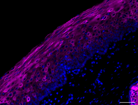
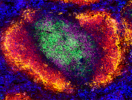
Immunocytochemistry (ICC) is characterized by imaging primary cells or cell lines in culture. Immunohistochemistry (IHC) refers to the detection of antibodies in tissue sections. Both ICC and IHC applications can utilize chromogenic or fluorescent detection methods. On our product technical data sheets, we use IHC-P to indicate that the antibody is useful in formalin-fixed paraffin-embedded (FFPE) sections and IHC-F to indicate the antibody is useful in tissue that has been fixed and frozen prior to sectioning. If there is only an IHC designation, check the literature citations to determine the method of tissue preparation compatible with each reagent.


View our IHC and ICC protocol videos for step-by-step guides to different microscopy workflows.

Newly Validated IHC Antibodies

Our scientists are continuously working to update application validation to our extensive clone library. Explore our newly validated IHC antibodies for cell and tissue imaging to drive discovery in various research fields, including oncology, immunology, and neurobiology.


View our recent antibody clones validated for IHC.

Fluorophores for Microscopy


Fluorescent detection allows visualization of multiple markers at a time, most commonly through the use of discrete excitation sources optimal for each fluorophore. Fluorescent detection introduces the opportunity for advanced imaging applications as well, like live-cell imaging, multiphoton imaging, super-resolution microscopy, FLIM, and FRET.


Although sensitivity can be a limitation of fluorescence microscopy at certain wavelengths, especially reagents that emit in the range of 350-450 nm due to increased autofluorescence of the sample, improved signal-to-noise can be obtained through varying enzymatic and immunologic amplification techniques, the use of higher sensitivity instrumentation, and near-infrared emitting fluorophores.


Right: Paraffin-embedded human prostate tissue was stained with anti-CD44 Alexa Fluor® 594 (red), purified anti-human CD107b (H4B4) antibody followed by anti-mouse IgG Alexa Fluor® 488 secondary antibody (green) and DAPI (blue).

We provide a vast selection of fluorophore antibody conjugates for your microscopy research. The table below highlights some of these conjugates and their emission profiles.


Direct Conjugate

Excitation max (nm)

Emission max (nm)

Emission color

DyLight™ 405

400

420

Blue

Brilliant Violet 421™

405

421

Blue

Brilliant Violet 510™

405

510

Green

DyLight™ 488

493

518

Green

Alexa Fluor® 488

495

519

Green

Alexa Fluor® 555

555

565

Orange

Spark YG™ 570

555

570

Orange

Alexa Fluor® 594

590

617

Red

DyLight™ 594

592

617

Red

Alexa Fluor® 647

650

665

Near-IR*

Alexa Fluor® 700

702

723

Near-IR*

* Human vision is not sensitive to light beyond ~650 nm; it is not possible to view near-IR fluorescent dyes.

Brilliant Violet™ Fluorophores


Brilliant Violet 421™  (BV421™) is used in the “blue” channel which is typically occupied by DAPI or Alexa Fluor® 405. Brilliant Violet 510™ (BV510™) is also excited at 405 nm, but emits at 510 nm. When your filter set-up is optimized, these organic polymers, BV421™ and BV510 ™, can be used simultaneously as bright, photostable options for multicolor microscopy.


Learn more about Brilliant Violet™ > 

Spark YG™ 570


Ideal for building multicolor microscopy panels, Spark YG™ 570 stands out as a reliable addition to any multicolor microscopy panel. With an excitation max of 555 nm and an emission max of 570 nm, it can be imaged using the filter sets commonly used for Alexa Fluor® 555, Cy3, or TRITC in either widefield or confocal microscopy. 


View our Spark YG™ 570 Products >

Five-Color Fluorescence Microscopy


With increased fluorophore options and the abundance of directly conjugated antibodies, it is possible to use fluorescence microscopy to look at 5 different markers on a single sample. For larger microscopy panels, it is especially critical to know your microscope and be sure it has the appropriate lasers and filters to capture the emission and excitation spectra of each of these distinct fluorophores.


We stained frozen C57BL/6 mouse spleen tissue using antibodies against CD4 and CD8a to detect T cells, B220 to stain B cells, and CD169 and F4/80 to detect tissue-resident macrophages. For this staining, we used antibodies directly-conjugated to bright photostable fluorophores including the Brilliant Violet™ and Alexa Fluor® dyes.

fullstaining

Microscope Excitation and Emission Filters


By optimizing your filter set-up to allow for the detection of Brilliant Violet 421™ and Brilliant Violet 510™, you can expand your microscopy panels. See how we optimized our set-up below.


Purified and Biotin-Labeled Primary Antibodies


Purified antibodies are the common go-to reagents for single color staining or in cases where secondary antibodies are desired for amplification. For amplification or multicolor applications, biotinylated antibodies are also effective tools. We provide hundreds of purified antibody products and dozens of biotinylated antibodies, quality-tested for ICC or IHC.


View our Purified Antibodies and Biotinylated Antibodies.


Secondary Reagents


Signal amplification is often required in imaging applications for lowly expressed antigens. One way to increase the likelihood of success when imaging a target is to amplify the primary signal without raising the background signal. In addition, amplifying a signal can also decrease the amount of exposure time, limiting the spillover of other fluors into your channel of interest. For these purposes, we provide a selection of dye-conjugated secondary reagents.


Explore our Secondary Reagents and Streptavidin Conjugates.


Chemical Probes


In addition to primary antibodies, a number of dyes or probes can be used to specifically stain subcellular structures. These can be used across a wide variety of microscopy applications, including staining of the nucleus, organelles, or cytoplasmic structures, such as actin and microtubules.


Our table highlights a number of our non-antibody chemical probes, their subcellular localization, and emission profiles.


Chemical Probe

Subcellular Localization

Excitation Max (nm)

Emission Max (nm)

Emission Color

Tag-it Violet™

Cytoplasm

405

450

Blue

DAPI

Nucleus

360

460

Blue

Helix NP™ Blue

Nucleus

430

470

Blue

MitoSpy™ Green FM

Mitochondria

490

516

Green

Helix NP™ Green

Nucleus

495

519

Green

Flash Phalloidin™ Green 488

Actin

488

520

Green

Apotracker™ Green

Phosphatidylserine

500

520

Green

Calcein Red-AM

Cytoplasm

560

574

Orange

MitoSpy™ Orange CMTMRos

Mitochondria

551

576

Orange

MitoSpy™ Red CMXRos

Mitochondria

577

598

Red

Flash Phalloidin™ Red 594

Actin

590

611

Red

MitoSpy™ NIR DilC1(5)

Mitochondria

638

658

Near-IR

Helix NP™ NIR

Nucleus

640

660

Near-IR

DRAQ5™

Nucleus

633

695

Near-IR


Reagents for Chromogenic Detection


Chromogenic detection methods are advantageous because a signal can be amplified simply by extending the amount of time and substrate in the reaction. Also, it does not require sophisticated instruments for detection, only a microscope with phase contrast. HRP detection can, however, be accompanied by endogenous background associated with cellular peroxidase activity, non-specific signal, and is only typically used to image a single marker at a time.


These methods have a long history in histology and pathology applications. Commonly used in chromogenic IHC are antibodies or streptavidin covalently attached with HRP or AlkPhos, that convert a substrate like DAB, AEC, or BCIP/NBT. These enzymes catalyze their substrates, leaving a deposit of color where the antibody has attached to the cell or tissue. We provide a variety of accessory reagents for performing IHC including, Ultra-Streptavidin (USA) HRP Detection Kits, and ACUITYAdvanced Biotin Free Polymer Detection Kits.


IHC staining of anti-DJ-1 antibody (PARK7) on FFPE normal (left) and Parkinson’s disease (right) brain tissue. Ultra Streptavidin (USA) HRP Detection Kit was used for detection followed by hematoxylin and bluing solution counterstaining. Scale bar: 50 µm.

ProductsHere

Login / Register
Remember me
Forgot your password? Reset password?
Create an Account