 Login / Register


- Clone
- C-11 (See other available formats)
- Regulatory Status
- RUO
- Other Names
- Cytokeratin, Keratin
- Isotype
- Mouse IgG1, κ
- Ave. Rating
- Submit a Review
- Product Citations
- publications

-

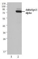
Whole cell extracts (15 µg protein) from Jurkat (negative control) and HeLa cells were resolved on a 4-20% Tris-glycine gel, transferred to a nitrocellulose membrane and probed with 0.5 µg/mL (1:1000 dilution) of Purified anti-Cytokeratin (pan reactive) Antibody, clone C-11, overnight at 4°C. Proteins were visualized by chemiluminescence detection HRP goat anti-mouse IgG Antibody (Cat. No. 405306) at a 1:3000 dilution. Direct-Blot™ HRP anti-β-Actin Antibody (Cat. No. 664804) was used as a loading control at a 1:25000 dilution (lower). Lane M: Molecular Weight marker. -

MCF-7 cells were stained with purified anti- Cytokeratin (pan reactive) (C-11) antibody, followed by staining with DyLight™ 594 conjugated goat anti-mouse IgG (red) antibody. Nuclei were stained with DAPI (blue). -

Hela cells stained with purified mouse monoclonal antibody against pan Cytokeratin (clone C-11), followed by Rhodamine Red-X conjugated Donkey anti-mouse IgG and DAPI.
| Cat # | Size | Price | Quantity Check Availability | Save | ||
|---|---|---|---|---|---|---|
| 628601 | 25 µg | 81€ | ||||
| 628602 | 100 µg | 175€ | ||||
Cytokeratins are intermediate filament proteins that are widely expressed in many tissues. These proteins are vital components of the cytoskeleton and interact with a number of other proteins. Cytokeratins are usually found in the cytoplasm,although some cytokeratins can be found in the nucleus and the nucleolus.
Product DetailsProduct Details
- Verified Reactivity
- Human
- Antibody Type
- Monoclonal
- Host Species
- Mouse
- Immunogen
- Keratin enriched fraction from human epidermoid carcinoma.
- Formulation
- This antibody is provided in phosphate-buffered solution, pH 7.2, containing 0.09% sodium azide at 0.5 mg/ml.
- Preparation
- The antibody was purified by affinity chromatography.
- Concentration
- 0.5 mg/ml
- Storage & Handling
- The antibody solution should be stored undiluted between 2°C and 8°C.
- Application
-
WB - Quality tested
ICC - Verified
IP, IHC-P - Reported in the literature, not verified in house - Recommended Usage
-
Each lot of this antibody is quality control tested by Western blotting. Western blotting, suggested working dilution(s): Use 5 µg antibody per 5 ml antibody dilution buffer for each mini-gel. For immunocytochemistry, a concentration range of 1.0 - 4.0 μg/ml is recommended. It is recommended that the reagent be titrated for optimal performance for each application.
- Application Notes
-
The C-11 monoclonal antibody reacts with a conserved epitope of human cytokeratin 4, 5, 6, 8, 10, 13, and 18. This antibody has been shown to be useful for Western blotting and has also been reported to be useful for immunoprecipitation, immunohistochemistry (paraffin sections), immunocytochemistry, and spatial biology (IBEX)7,8.
-
Application References
(PubMed link indicates BioLegend citation) -
- Kovarik J. 1988. Int. J. Cancer Suppl. 3:50.
- Bartek J, et al. 1991. J. Pathol. 164:215.
- Chernyavsky A I, 2007. J. Biol. Chem. doi:10.1074/jbc.M611365200. PubMed
- Comito G, et al. 2012. Cancer Lett. 324:31. PubMed
- Vojt-òsek B, et al. 1989. Folia Biol (Praha). 35:373. (reactivity with rat, dog, sheep, pig, cow)
- Dekaney CM, et al. 2005. Gastroenterology 129:1567. (ICC)
- Radtke AJ, et al. 2020. Proc Natl Acad Sci U S A. 117:33455-65. (SB) PubMed
- Radtke AJ, et al. 2022. Nat Protoc. 17:378-401. (SB) PubMed
- Product Citations
-
- RRID
-
AB_439774 (BioLegend Cat. No. 628601)
AB_439775 (BioLegend Cat. No. 628602)
Antigen Details
- Structure
- Intermediate filament protein, contains three coiled-coil domains. Cytokeratins exist as heterotetramers composed of two type I and two type II keratin subunits
- Distribution
-
Cytokeratins are widely expressed proteins usually found in the cytoplasm although some cytokeratins can also be found in the nucleus and the nucleolus
- Function
- Intermediate filament protein involved with the cytoskeleton.
- Interaction
- Most cytokeratins interact with a number of other proteins such as 14-3-3, EGF receptor, and many others
- Biology Area
- Cell Biology, Cell Motility/Cytoskeleton/Structure, Neuroscience, Neuroscience Cell Markers
- Molecular Family
- Intermediate Filaments
- Gene ID
- 3875 View all products for this Gene ID
- UniProt
- View information about Cytokeratin pan reactive on UniProt.org

Related FAQs
Other Formats
View All Cytokeratin Reagents Request Custom Conjugation| Description | Clone | Applications |
|---|---|---|
| Purified anti-Cytokeratin (pan reactive) | C-11 | WB,ICC,IP,IHC-P |
| Alexa Fluor® 647 anti-Cytokeratin (pan reactive) | C-11 | ICC,SB,ICFC |
| Alexa Fluor® 594 anti-Cytokeratin (pan reactive) | C-11 | ICC |
| Alexa Fluor® 488 anti-Cytokeratin (pan reactive) | C-11 | ICC,IHC-P |
Customers Also Purchased
Compare Data Across All Formats
This data display is provided for general comparisons between formats.
Your actual data may vary due to variations in samples, target cells, instruments and their settings, staining conditions, and other factors.
If you need assistance with selecting the best format contact our expert technical support team.
-
Purified anti-Cytokeratin (pan reactive)

Whole cell extracts (15 µg protein) from Jurkat (negative co... 
MCF-7 cells were stained with purified anti- Cytokeratin (pa... 
Hela cells stained with purified mouse monoclonal antibody a... -
Alexa Fluor® 647 anti-Cytokeratin (pan reactive)

MCF-7 cells were fixed with 1% paraformaldehyde (PFA) for 10... 
Confocal image of C57BL/6 mouse thymus sample acquired using... 
MCF7 cells were fixed and permeabilized using Cyto-Fast Fix/... 
MCF7 cells (positive control, solid histogram), and Jurkat c... -
Alexa Fluor® 594 anti-Cytokeratin (pan reactive)

MCF-7 cells were fixed with 1% paraformaldehyde (PFA) for te... -
Alexa Fluor® 488 anti-Cytokeratin (pan reactive)

MCF-7 mammary adenocarcinoma cells were fixed, permeabilized... 
Human paraffin-embedded colon tissue slices were prepared wi...


Follow Us