Ubiquitin as an IHC Marker for Neurodegeneration


Ubiquitin is a small regulatory protein that is expressed in all eukaryotic cells. Ubiquitination, a process whereby ubiquitin is attached to a substrate protein, is involved in many cellular processes including protein degradation, endocytic trafficking, antigen processing, organelle biogenesis, transcriptional regulation, and DNA repair. Aberrant ubiquitination and/or dysfunction in ubiquitin-mediated pathways have been linked to numerous disorders.


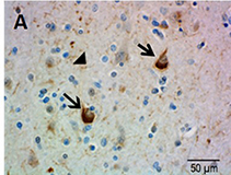
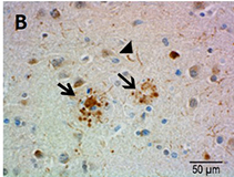
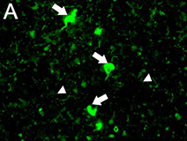
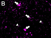
Ubiquitin immunostaining is often among the basic immunohistochemical (IHC) panels for neurodegeneration detection. Ubiquitin accumulates in morphologically distinct deposits associated with several neurodegenerative disorders, such as Tauopathies, Alzheimer’s (AD) and Parkinson’s (PD) diseases. These disorders are collectively called proteinopathies and share a common feature: abnormal misfolding of key proteins that increases their tendency to self-aggregate and form proteinaceous inclusions. Loss of normal function, resistance to degradation and clearance, and interference with normal cellular processes are attributed to the aggregated state of proteins involved.


Antibodies against ubiquitin can visualize the presence of disease-related structures, such as cytoplasmic or extracellular inclusions. The use of disease specific antigens or biomarkers can supplement data obtained from ubiquitin staining to further distinguish among pathologies. Some examples are highlighted in the table below.

Disease features detected by ubiquitin staining:


Disease Pathology Complementary staining
Synucleinopathies (e.g. PD, multiple system atrophy)
  • Intracytoplasmic α-Synuclein inclusions known as Lewy bodies
  • Abnormal α-Synuclein filaments found in Lewy neurites
α-Synuclein
Alzheimer’s disease
  • Extracellular, amyloid beta (Aβ)-containing deposits known as amyloid or neuritic plaques
  • Neurofibrillary tangles composed of aggregated hyper-phosphorylated Tau
  • Abnormal neuronal processes known as dystrophic neurites often associated with neuritic plaques
Aβ, Tau
Frontotemporal Lobar Degeneration (FTLD)-TDP
  • Misfolded TDP-43 detected in neuronal nuclear and cytoplasmic inclusions and neurites.
TDP-43

Advantages of using ubiquitin as a marker for neurodegeneration:


  • Ubiquitin staining for microscopy is reliable and reproducible
  • Protocol is easier and less labor-intensive compared to other histological techniques, such as silver impregnation stains
  • Simplifies evaluation of and distinction between diseased and non-diseased brains
  • Allows for easy assessment of inclusion density (e.g. amyloid plaques or Tau tangles)
  • Widespread use of IHC makes it a highly accessible technique


Fig 1. IHC staining of FFPE AD brain tissue stained with anti-Ubiquitin antibody, clone P4G7. Arrows point to Tau tangles (A) and neuritic plaques (B). Arrowheads point to neuropil threads in A & B.


Fig 2. IHC staining of FFPE AD brain tissue co-stained with (A) anti-Tau  phospho (Ser396) (clone PHF-13, green) and (B) anti-Ubiquitin (clone P4G7, magenta) antibodies. (C) Nuclei were counterstained with DAPI (blue). (D) Three-channel merge is shown. Arrows and arrowheads point to Tau tangles and neuropil threads, respectively.

ProductsHere

Login / Register
Remember me
Forgot your password? Reset password?
Create an Account