 Login / Register


- Clone
- A20 (See other available formats)
- Regulatory Status
- RUO
- Other Names
- T200, Ly-5.1, LCA
- Isotype
- Mouse (A.SW) IgG2a, κ
- Ave. Rating
- Submit a Review
- Product Citations
- publications

-

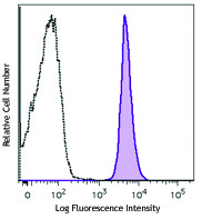
SJL mouse splenocytes were stained with CD45.1 (clone A20) Brilliant Violet 510™ (filled histogram) or mouse IgG2a Brilliant Violet 510™ isotype control (open histogram).
| Cat # | Size | Price | Quantity Check Availability | Save | ||
|---|---|---|---|---|---|---|
| 110741 | 50 µg | 219€ | ||||
CD45.1 is an alloantigen of CD45, expressed by Ly5.1 bearing mouse strains (e.g., RIII, SJL/J, STS/A, DA). CD45, a member of the protein tyrosine phosphatase (PTP) family, is a 180-240 kD glycoprotein expressed on all hematopoietic cells except mature erythrocytes and platelets. There are multiple isoforms in mice that play key roles in TCR and BCR signal transduction. These isoforms are very specific to the activation and maturation states of the cell as well as specific cell types. The primary ligands for CD45 are galectin-1, CD2, CD3, CD4, TCR, CD22, and Thy-1.
Product DetailsProduct Details
- Verified Reactivity
- Mouse
- Antibody Type
- Monoclonal
- Host Species
- Mouse
- Immunogen
- SJL mouse thymocytes and splenocytes
- Formulation
- Phosphate-buffered solution, pH 7.2, containing 0.09% sodium azide and BSA (origin USA).
- Preparation
- The antibody was purified by affinity chromatography and conjugated with Brilliant Violet 510™ under optimal conditions.
- Storage & Handling
- The antibody solution should be stored undiluted between 2°C and 8°C, and protected from prolonged exposure to light. Do not freeze.
- Application
-
FC - Quality tested
- Recommended Usage
-
Each lot of this antibody is quality control tested by immunofluorescent staining with flow cytometric analysis. For flow cytometric staining, the suggested use of this reagent is ≤0.5 µg per million cells in 100 µl volume. It is recommended that the reagent be titrated for optimal performance for each application.
Brilliant Violet 510™ excites at 405 nm and emits at 510 nm. The bandpass filter 510/50 nm is recommended for detection, although filter optimization may be required depending on other fluorophores used. Be sure to verify that your cytometer configuration and software setup are appropriate for detecting this channel. Refer to your instrument manual or manufacturer for support. Brilliant Violet 510™ is a trademark of Sirigen Group Ltd.
Learn more about Brilliant Violet™.
This product is subject to proprietary rights of Sirigen Inc. and is made and sold under license from Sirigen Inc. The purchase of this product conveys to the buyer a non-transferable right to use the purchased product for research purposes only. This product may not be resold or incorporated in any manner into another product for resale. Any use for therapeutics or diagnostics is strictly prohibited. This product is covered by U.S. Patent(s), pending patent applications and foreign equivalents. - Excitation Laser
-
Violet Laser (405 nm)
- Application Notes
-
The A20 antibody does not react with leukocytes or mouse cells expressing the CD45.2 alloantigen. Additional reported applications (for relevant formats of this clone) include: immunoprecipitation3, in vitro blocking of B cell responses1,2, immunohistochemical staining of frozen sections: OCT embedded7 and acetone-fixed4-6 (direct immunofluorescence detection with fluorochrome conjugated A20 was used in (5) and (6)).
- Application References
-
- Yakura H, et al. 1983. J. Exp. Med. 157:1077. (Block)
- Yakura H, et al. 1986. J. Immunol. 136:2729. (Block)
- Shen FW, et al. 1986. Immunogenetics 24:146. (IP)
- Suzuki K, et al. 2000. Immunity 13:691. (IHC-F)
- Werner N, et al. 2002. Arterioscler. Thromb. Vasc. Biol. 22:1567. (IHC-F)
- Lessner SM, et al. 2002. Am. J. Pathol. 160:2145. (FC, IHC-F)
- Chen CC, et al. 2005. P. Natl. Acad. Sci. USA 102:11408 (IHC-F)
- Pascal V, et al. 2007. J. Immunol. 179:1751. (FC)
- Mende I, et al. 2006. Blood 107:1383. (IHC-F, FC)
- Phan TG, et al. 2007. Nature Immunol. 8:992. (FC)
- Wither DR, et al. 2009. J. Immunol. 183:5079. PubMed
- Pascal V, et al.2007. J. Immunol. 179:1751. PubMed
- Lee SW, et al. 2009. J. Immunol. 182:6753. PubMed
- Takada K, et al. 2009. J. Exp Med. 206:2253. PubMed
- Beamer CA, et al. 2007. Am. J. Respir. Cell. Mol. Biol. 37:729. (FC) PubMed
- Li LX, et al. 2010. J. Immunol. 184:1728. PubMed
- Hosoi A, et al. 2008. Cancer Res. 68:3941. (FC) PubMed
- Kenna TJ, et al. 2008. Blood 111:2091. PubMed
- Kohlmeier JE, et al. 2008. Immunity. 29:101. (FC) PubMed
- Product Citations
-
- RRID
-
AB_2563378 (BioLegend Cat. No. 110741)
Antigen Details
- Structure
- Protein tyrosine phosphatase (PTP) family, 180-240 kD
- Distribution
-
All hematopoietic cells except mature erythrocytes and platelets of the CD45.1 strain of mice
- Function
- Phosphatase, T and B cell activation
- Ligand/Receptor
- Galectin-1, CD2, CD3, CD4
- Biology Area
- Cell Biology, Immunology, Inhibitory Molecules, Neuroscience, Neuroscience Cell Markers
- Molecular Family
- CD Molecules
- Antigen References
-
1. Barclay A, et al. 1997. The Leukocyte Antigen FactsBook Academic Press.
2. Trowbridge IS, et al. 1993. Annu. Rev. Immunol. 12:85.
3. Kishihara K, et al. 1993. Cell 74:143.
4. Pulido R, et al. 1988. J. Immunol. 140:3851. - Gene ID
- 19264 View all products for this Gene ID
- UniProt
- View information about CD45.1 on UniProt.org


Related FAQs
Other Formats
View All CD45.1 Reagents Request Custom ConjugationCustomers Also Purchased

Compare Data Across All Formats
This data display is provided for general comparisons between formats.
Your actual data may vary due to variations in samples, target cells, instruments and their settings, staining conditions, and other factors.
If you need assistance with selecting the best format contact our expert technical support team.
-
Biotin anti-mouse CD45.1

SJL mouse splenocytes stained with biotinylated A20, followe... -
FITC anti-mouse CD45.1

SJL mouse splenocytes stained with A20 FITC -
PE anti-mouse CD45.1

SJL (solid line) and C57BL/6 (broken line) splenocytes stain... -
Purified anti-mouse CD45.1

SJL mouse splenocytes stained with purified A20, followed by... -
APC anti-mouse CD45.1

SJL mouse splenocytes stained with A20 APC -
APC/Cyanine7 anti-mouse CD45.1

SJL mouse splenocytes were stained with CD45.1 (clone A20) A... -
Alexa Fluor® 488 anti-mouse CD45.1

SJL mouse splenocytes stained with A20 Alexa Fluor® 488 -
Alexa Fluor® 647 anti-mouse CD45.1

SJL mouse splenocytes stained with A20 Alexa Fluor® 647 -
Pacific Blue™ anti-mouse CD45.1

SJL mouse splenocytes stained with A20 Pacific Blue™ -
Alexa Fluor® 700 anti-mouse CD45.1

SJL mouse splenocytes stained with A20 Alexa Fluor® 700 -
Brilliant Violet 650™ anti-mouse CD45.1

SJL mouse splenocytes were stained with CD45.1 (clone A20) B... -
PerCP anti-mouse CD45.1

SJL mouse splenocytes stained with A20 PerCP -
PerCP/Cyanine5.5 anti-mouse CD45.1

SJL mouse splenocytes stained with A20 PerCP/Cyanine5.5 -
PE/Cyanine7 anti-mouse CD45.1

SJL splenocytes stained with A20 PE/Cyanine7 -
Brilliant Violet 421™ anti-mouse CD45.1

SJL mouse splenocytes were stained with CD45.1 (clone A20) B... 
C57BL/6 mouse splenocytes were stained with CD45.1 (clone A2... -
Brilliant Violet 570™ anti-mouse CD45.1

SJL mouse splenocytes were stained with CD45.1 (clone A20) B... -
Brilliant Violet 605™ anti-mouse CD45.1

SJL mouse splenocytes were stained with CD45.1 (clone A20) B... -
Brilliant Violet 711™ anti-mouse CD45.1

SJL mouse splenocytes were stained with CD45.1 (clone A20) B... -
Brilliant Violet 510™ anti-mouse CD45.1

SJL mouse splenocytes were stained with CD45.1 (clone A20) B... -
Brilliant Violet 785™ anti-mouse CD45.1

SJL mouse splenocytes were stained with CD45.1 (clone A20) B... -
Purified anti-mouse CD45.1 (Maxpar® Ready)

NOD (red) and C57BL/6 (blue) mouse splenocytes stained with ... -
PE/Dazzle™ 594 anti-mouse CD45.1

SJL mouse splenocytes were stained with CD45.1 (clone A20) P... -
Alexa Fluor® 594 anti-mouse CD45.1

SJL mouse frozen spleen section was fixed with 4% paraformal... -
APC/Fire™ 750 anti-mouse CD45.1

SJL mouse splenocytes were stained with CD45.1 (clone A20) A... -
TotalSeq™-A0178 anti-mouse CD45.1
-
TotalSeq™-B0178 anti-mouse CD45.1
-
TotalSeq™-C0178 anti-mouse CD45.1
-
Spark UV™ 387 anti-mouse CD45.1

SJL mouse splenocytes were surface stained with anti-mouse C... -
Spark Blue™ 550 anti-mouse CD45.1 (Flexi-Fluor™)
-
Spark Red™ 718 anti-mouse CD45.1 (Flexi-Fluor™)


Follow Us