 Login / Register


- Clone
- 9E10 (See other available formats)
- Regulatory Status
- RUO
- Other Names
- myc tag, c-Myc tag, cMyc tag, Myc epitope tag
- Isotype
- Mouse IgG1, κ
- Ave. Rating
- Submit a Review
- Product Citations
- publications

-

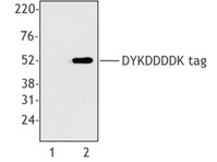
Total cell lysates (15 µg protein) from 293E cells transfected with Multi-tag (lane 1) or non-transfected cells (lane 2) were resolved by electrophoresis (4-12% Bis-Tris gel), transferred to nitrocellulose, and probed with 1 µg/mL purified anti-c-Myc antibody (1:500 dilution), clone 9E10 (upper blot). Proteins were visualized by chemiluminescence detection using a 1:3000 diluted goat anti-mouse-IgG secondary antibody conjugated to HRP for the anti-c-Myc antibody or 1:5000 diluted Direct-Blot HRP anti-β-Actin antibody, clone 2F1-1 (lower blot). Lane M: Molecular weight ladder. -

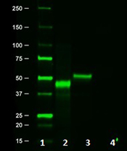
Whole cell extracts (15 µg total protein) from HeLa cells mixed with the indicated amount of recombinant Posi-tag epitope tag protein (Cat. No. 931301) were resolved by 4-12% Bis-Tris gel electrophoresis, transferred to a PVDF membrane, and probed with 1.0 µg/mL purified anti-c-Myc antibody (clone 9E10) overnight at 4°C. Proteins were visualized by chemiluminescence detection using HRP goat anti-mouse IgG antibody at a 1:3000 dilution. Direct-Blot™ HRP anti-GAPDH antibody (Cat. No. 607904) was used as a loading control at a 1:25000 dilution (lower). Lane M: Molecular weight marker. -

Whole cell extracts (250 µg total protein) prepared from HeLa cells mock-transfected (-) or transfected (+) with multi-tag plasmid containing c-Myc epitope sequence were immunoprecipitated overnight with 2.5 µg of purified mouse IgG1, κ isotype ctrl antibody (Cat. No. 400101) or purified anti-c-Myc antibody (clone 9E10). The resulting IP fractions and whole cell extract input (6%) were resolved by 4-12% Bis-Tris gel electrophoresis, transferred to a PVDF membrane and probed with purified anti-c-Myc antibody (clone 9E11) at 1.0 µg/mL (1:1000 dilution). Lane M: Molecular weight marker. -

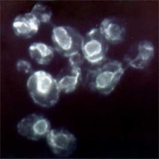
ICC staining of purified anti-c-Myc antibody (clone 9E10) on HeLa cells transfected with multi-tag plasmid containing c-Myc epitope sequence. The cells were fixed with 4% PFA, permeabilized with a buffer containing 0.1% Triton X-100, and blocked with 5% fetal bovine serum in PBS for 1 hour. The cells were then incubated with 1 µg/mL of the primary antibody overnight at 4°C, followed by incubation with 2.5 µg/mL of Alexa Fluor® 488 goat anti-mouse IgG antibody (green) for one hour at room temperature. Nuclei were counterstained with DAPI (blue), and the slide was mounted with ProLong™ Gold Antifade Mountant. The image was captured with a 40X objective. Scale bar: 100 µm -

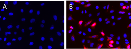
IHC staining of anti-c-Myc antibody (clone 9E10) on formalin-fixed paraffin-embedded human bladder tissue. Following antigen retrieval using 0.1M Tris-buffered saline w/ Tween 20 (Cat. No. 925501), the tissue was incubated with 1 µg/mL of the primary antibody overnight at 4°C, followed by incubation with 2.5 µg/mL of Alexa Fluor® 647 goat anti-mouse IgG antibody for one hour at room temperature. Nuclei were counterstained with DAPI, and the slide was mounted with ProLong™ Gold Antifade Mountant. The image was captured with a 20x objective. Scale bar: 100 µm
| Cat # | Size | Price | Quantity Check Availability | Save | ||
|---|---|---|---|---|---|---|
| 626801 | 25 µg | 81€ | ||||
| 626802 | 100 µg | 175€ | ||||
The 9E10 monoclonal antibody recognizes the amino acid sequence EQKLISEEDL, which is a specific portion of the human c-myc gene product. This antibody is highly specific and is unlikely to alter the activity of the cloned sequence.
Product DetailsProduct Details
- Verified Reactivity
- Epitope tag
- Reported Reactivity
- Species independent
- Antibody Type
- Monoclonal
- Host Species
- Mouse
- Immunogen
- Amino acids 408-439, C-terminal region of human c-myc
- Formulation
- This antibody is provided in phosphate-buffered solution, pH 7.2, containing 0.09% sodium azide at 0.5 mg/ml.
- Preparation
- The antibody was purified by affinity chromatography.
- Concentration
- 0.5 mg/mL
- Storage & Handling
- Upon receipt, store undiluted between 2°C and 8°C.
- Application
-
WB - Quality tested
IP, ICC, IHC-P - Verified
Purification - Reported in the literature, not verified in house - Recommended Usage
-
Each lot of this antibody is quality control tested by Western blotting. For Western blotting, the suggested use of this reagent is 0.5 - 1.0 µg per ml. For immunoprecipitation, the suggested use of this reagent is 2.5 µg/test. For immunocytochemistry, a concentration range of 1.0 - 2.5 μg/mL is recommended. For immunohistochemistry on formalin-fixed paraffin-embedded tissue sections, a concentration range of 1.0 - 5.0 µg/ml is suggested. It is recommended that the reagent be titrated for optimal performance for each application.
- Application Notes
-
*Our Posi-Tag Control Protein (931301) can be used as a helpful positive control for this antibody.
- Additional Product Notes
-
This antibody recognizes endogenous human c-Myc protein in IHC-P. Reactivity with endogenous c-Myc in other applications is not verified.
- Application References
-
- Rao H, et al. 2001. Nature. 410:955.
- Peters CS, et al. 2001. J Biol Chem. 276:13718.
- Munro S, Pelham HR. 1987. Cell. 48:899.
- Evan GI, et al. 1985. Mol Cell Biol. 5:3610.
- Product Citations
-
- RRID
-
AB_2235686 (BioLegend Cat. No. 626801)
AB_2148451 (BioLegend Cat. No. 626802)
Antigen Details
- Biology Area
- Cell Biology, Protein Purification, Protein Synthesis
- Antigen References
-
- Adams JM, et al. 1983. Proc. Natl. Acad. Sci. USA 80:1982.
- Atchley WR, et al. 1995. Proc. Natl. Acad. Sci. USA 92:10217.
- Battey J, et al. 1983. Cell 34:779.
- Beimling P, et al. 1985. Biochemistry 24:6349.
- Gene ID
- 4609 View all products for this Gene ID
- UniProt
- View information about c-Myc on UniProt.org

Related FAQs
Other Formats
View All c-Myc Reagents Request Custom Conjugation| Description | Clone | Applications |
|---|---|---|
| Purified anti-c-Myc | 9E10 | WB,ICC,IP,IHC-P,Purification |
| Anti-c-Myc Tag (9E10) Affinity Gel | 9E10 | Purification,IP |
| Biotin anti-c-Myc | 9E10 | WB,ICC |
| Direct-Blot™ HRP anti-c-Myc | 9E10 | WB |
| Alexa Fluor® 594 anti-c-Myc | 9E10 | ICC |
| Ultra-LEAF™ Purified anti-c-Myc | 9E10 | WB,ELISA,ICC,IP,IHC-P,Purification |
| Alexa Fluor® 647 anti-c-Myc | 9E10 | ICC |
| Alexa Fluor® 488 anti-c-Myc Antibody | 9E10 | ICC |
Customers Also Purchased
Compare Data Across All Formats
This data display is provided for general comparisons between formats.
Your actual data may vary due to variations in samples, target cells, instruments and their settings, staining conditions, and other factors.
If you need assistance with selecting the best format contact our expert technical support team.
-
Purified anti-c-Myc

Total cell lysates (15 µg protein) from 293E cells transfect... 
Whole cell extracts (15 µg total protein) from HeLa cells mi... 
Whole cell extracts (250 µg total protein) prepared from HeL... 
ICC staining of purified anti-c-Myc antibody (clone 9E10) on... 
IHC staining of anti-c-Myc antibody (clone 9E10) on formalin... -
Anti-c-Myc Tag (9E10) Affinity Gel

Binding capacity of anti-c-Myc agarose for C-terminal c-Myc ... 
Stable activity of anti-c-Myc agarose over ten rounds of aff... -
Biotin anti-c-Myc

Western blot analysis of 1, 0.5, 0.1 µl (lanes 1, 2, 3 respe... 
Immunofluorescence of Myc-tagged plasmid-transfected A549 ce... -
Direct-Blot™ HRP anti-c-Myc

Recombinant Posi-Tag Epitope Tag Protein (1 µL, diluted 1:10... -
Alexa Fluor® 594 anti-c-Myc

HeLa cells transiently transfected with c-Myc tag fused prot... 
HeLa cells transiently transfected with c-Myc tag fused prot... -
Ultra-LEAF™ Purified anti-c-Myc
Total cell lysates (15 µg protein) from 293E cells transfect... -
Alexa Fluor® 647 anti-c-Myc

Untransfected (panel A) or multi-tag transfected (panel B) H... -
Alexa Fluor® 488 anti-c-Myc Antibody

Untransfected (panel A) or multi-tag transfected (panel B) H...


Follow Us