 Login / Register


- Clone
- WL4G10 (See other available formats)
- Regulatory Status
- RUO
- Other Names
- CMD1A, HGPS, LGMD1B, LMN1, LMNL1, MADA, PRO1
- Isotype
- Mouse IgG1, κ
- Ave. Rating
- Submit a Review
- Product Citations
- publications

-

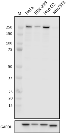
Total cell lysates (15 µg total protein) from the indicated cell lines were resolved by 4-12% Bis-Tris gel electrophoresis, transferred to a PVDF membrane, and probed with 1.0 µg/mL (1:500 dilution) of Purified anti-Lamin A/C Antibody, clone WL4G10, overnight at 4°C. Proteins were visualized by chemiluminescence detection using HRP goat anti-mouse IgG Antibody (Cat. No. 405306) at a 1:3000 dilution. Direct-Blot™ HRP anti-GAPDH Antibody (Cat. No. 607904) was used as a loading control at a 1:10000 dilution (lower). Lane M: Molecular Weight marker. -

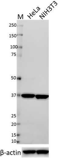
Total cell lysates (15 µg total protein) from Daudi (reduced expression control) and HeLa cells were resolved by 4-12% Bis-Tris gel electrophoresis, transferred to a PVDF membrane, and probed with 0.1 µg/mL (1:5000 dilution) of Purified anti-Lamin A/C Antibody, clone WL4G10, overnight at 4°C. Proteins were visualized by chemiluminescence detection using HRP goat anti-mouse IgG Antibody (Cat. No. 405306) at a 1:3000 dilution. Direct-Blot™ HRP anti-GAPDH Antibody (Cat. No. 607904) was used as a loading control at a 1:10000 dilution (lower). Lane M: Molecular Weight marker. Predicted LMNA expression data was obtained from Human Protein Atlas. -

HeLa cells were fixed with 4% paraformaldehyde for 10 minutes, permeabilized with Triton X-100 for 10 minutes, and blocked with 5% FBS for 60 minutes. Cells were then intracellularly stained with a 1:100 dilution (5 µg/mL) of either Purified Mouse IgG1, κ Isotype Control Antibody (Cat. No. 401402, panel A) or Purified anti-Lamin A/C Antibody, Clone WL4G10 (panel B) overnight at 4°C, followed by incubation with Alexa Fluor® 594 goat anti-mouse IgG (Cat. No. 405326) at 2.0 µg/mL. Nuclei were counterstained with DAPI and the image was captured with a 60X objective. -

Daudi cells (negative control, open histogram) or Jurkat cells (positive control, filled histogram) were fixed and permeabilized using True-Nuclear™ Transcription Factor Buffer Set (Cat. No. 424401) and intracellularly stained with purified anti-TAL1 (clone 4G8C07), or purified mouse IgG1, κ isotype control (open histogram, dashed line) (representative histogram for both cell lines) (Cat. No. 401401), followed by PE goat anti-mouse IgG (Cat. No. 405307).
| Cat # | Size | Price | Quantity Check Availability | Save | ||
|---|---|---|---|---|---|---|
| 600001 | 25 µg | 81€ | ||||
| 600002 | 100 µg | 203€ | ||||
Lamins A and C are structural components of the lamina, a scaffold of proteins localized to the nuclear inner membrane, and contribute to nuclear membrane dynamics and chromatin structure. Mutations in the LMNA gene have been linked to multiple laminopathy disorders, including Hutchinson-Gilford progeria syndrome (HGPS), an autosomal dominant disorder characterized by the appearance of accelerated and dramatic aging. Patients with HGPS have a mutation in the LMNA gene that results in aberrant splicing and production of the toxic, truncated Lamin A protein progerin that functions in a dominant negative pattern.
Product DetailsProduct Details
- Verified Reactivity
- Human
- Antibody Type
- Monoclonal
- Host Species
- Mouse
- Immunogen
- GST fused to C-terminal fragment of Lamin A (amino acid residues 422-664)
- Formulation
- Phosphate-buffered solution, pH 7.2, containing 0.09% sodium azide.
- Preparation
- The antibody was purified by affinity chromatography.
- Concentration
- 0.5 mg/mL
- Storage & Handling
- The antibody solution should be stored undiluted between 2°C and 8°C.
- Application
-
WB - Quality tested
ICC, ICFC - Verified - Recommended Usage
-
Each lot of this antibody is quality control tested by Western blotting. For Western blotting, the suggested use of this reagent is 0.1 - 1.0 µg per mL. For flow cytometric staining using our True-Phos™ Perm Buffer in Cell Suspensions Protocol, the suggested use of this reagent is ≤ 0.25 µg per million cells in 100 µL volume. It is recommended that the reagent be titrated for optimal performance for each application.
- Application Notes
-
WL4G10 does not recognize mouse Lamin A or C.
WL4G10 failed to immunoprecipitate Lamin A or C from HeLa lysates.
During PD testing for ICC applications, WL4G10 staining of PFA-fixed HeLa cells was compatible with both Methanol and Triton X-100 permeabilization methods; observed staining was slightly better with Triton X-100 permabilization. - RRID
-
AB_2810655 (BioLegend Cat. No. 600001)
AB_2810656 (BioLegend Cat. No. 600002)
Antigen Details
- Structure
- Lamin A is a 664 amino acid protein with a predicted molecular weight of 74 kD. Lamin C is a 572 amino acid protein with a predicted molecular weight of 65 kD.
- Distribution
-
Ubiquitously expressed/Nucleoplasmic side of inner nuclear membrane
- Function
- Fibrous component of nuclear lamina, provides framework for nuclear envelope, may interact with chromatin
- Cell Type
- Neurons
- Biology Area
- Cell Biology, Cell Motility/Cytoskeleton/Structure, Neuroscience, Neuroscience Cell Markers, Western Blot Controls
- Molecular Family
- Nuclear Markers, Organelle Markers
- Gene ID
- 4000 View all products for this Gene ID
- UniProt
- View information about Lamin A on UniProt.org

Related FAQs
Other Formats
View All Lamin A/C Reagents Request Custom Conjugation| Description | Clone | Applications |
|---|---|---|
| Purified anti-Lamin A/C | WL4G10 | WB,ICC,ICFC |
Customers Also Purchased
Compare Data Across All Formats
This data display is provided for general comparisons between formats.
Your actual data may vary due to variations in samples, target cells, instruments and their settings, staining conditions, and other factors.
If you need assistance with selecting the best format contact our expert technical support team.


Follow Us