 Login / Register


- Clone
- 8C5.5 (See other available formats)
- Regulatory Status
- RUO
- Other Names
- DSRED
- Isotype
- Mouse IgG2a, κ
- Ave. Rating
- Submit a Review
- Product Citations
- publications

-

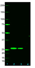
Total cell lysates (15 µg total protein) from mock-transfected 293E or 293E transfected with mCherry- fused protein were resolved by 4-12% Bis-Tris gel electrophoresis, transferred to a PVDF membrane, and probed with 0.25 µg/mL (1:2000 dilution) of Purified anti-mCherry Antibody, clone 8C5.5, overnight at 4°C. Proteins were visualized by chemiluminescence detection using HRP goat anti-mouse IgG Antibody (Cat. No. 405306) at a 1:3000 dilution. Direct-Blot™ HRP anti-GAPDH Antibody (Cat. No. 607904) was used as a loading control at a 1:1000 dilution (lower). Lane M: Molecular Weight marker. -

mCherry-transfected 293T cells were stained with purified anti-mCherry (clone 8C5.5) antibody, followed by staining with DyLight™ 488 Goat anti-mouse IgG antibody (green). Nuclei were counterstained with DAPI (blue). Non-transfected 293T cells showed nuclei staining only. The image was captured with a 40X objective.
| Cat # | Size | Price | Quantity Check Availability | Save | ||
|---|---|---|---|---|---|---|
| 677701 | 25 µg | 100€ | ||||
| 677702 | 100 µg | 249€ | ||||
mCherry is a red fluorescent protein that is often used to tag target proteins and to monitor their subcellular localization. It is the most widely used photostable protein in the class of red-shifted fluorescent proteins1,2. mCherry derives from proteins initially isolated from Cnidarians.
Product DetailsProduct Details
- Antibody Type
- Monoclonal
- Host Species
- Mouse
- Immunogen
- mCherry.
- Formulation
- Phosphate-buffered solution, pH 7.2, containing 0.09% sodium azide.
- Preparation
- The antibody was purified by affinity chromatography.
- Concentration
- 0.5 mg/ml
- Storage & Handling
- The antibody solution should be stored undiluted between 2°C and 8°C.
- Application
-
WB - Quality tested
ICC, IP - Verified - Recommended Usage
-
Each lot of this antibody is quality control tested by Western blotting. For Western blotting, the suggested use of this reagent is 1.0 - 2.5 µg per ml. For immunocytochemistry, a concentration range of 1.0 - 5.0 µg per ml is recommended. For immunoprecipitation, the suggested use of this reagent is 2.0 - 10 µg per ml. It is recommended that the reagent be titrated for optimal performance for each application.
- Product Citations
-
- RRID
-
AB_2801131 (BioLegend Cat. No. 677701)
AB_2565910 (BioLegend Cat. No. 677702)
Antigen Details
- Structure
- 236 amino acids with a predicted molecular weight of approximately 27 kD.
- Function
- Fluorescent protein.
- Biology Area
- Cell Biology
- Antigen References
-
1. Shaner NC, et al. 2008. Nat. Methods 5:545.
2. Shaner NC, et al. 2005. Nat. Methods 2:905. - Gene ID
- NA
- UniProt
- View information about mCherry on UniProt.org

Related FAQs
Other Formats
View All mCherry Reagents Request Custom Conjugation| Description | Clone | Applications |
|---|---|---|
| Purified anti-mCherry | 8C5.5 | WB,ICC,IP |
| Direct-Blot™ HRP anti-mCherry | 8C5.5 | WB |
Customers Also Purchased
Compare Data Across All Formats
This data display is provided for general comparisons between formats.
Your actual data may vary due to variations in samples, target cells, instruments and their settings, staining conditions, and other factors.
If you need assistance with selecting the best format contact our expert technical support team.
-
Purified anti-mCherry

Total cell lysates (15 µg total protein) from mock-transfect... 
mCherry-transfected 293T cells were stained with purified an... -
Direct-Blot™ HRP anti-mCherry

10 µg of total protein extract from 293E cells transfected ...


Follow Us