 Login / Register


- Clone
- SMI 99 (See other available formats)
- Regulatory Status
- RUO
- Other Names
- MBP, Golli-MBP, myelin A1 protein, myelin membrane encephalitogenic protein
- Previously
-
Covance Catalog# SMI-99P
- Isotype
- Mouse IgG2b, κ
- Ave. Rating
- Submit a Review
- Product Citations
- publications

-

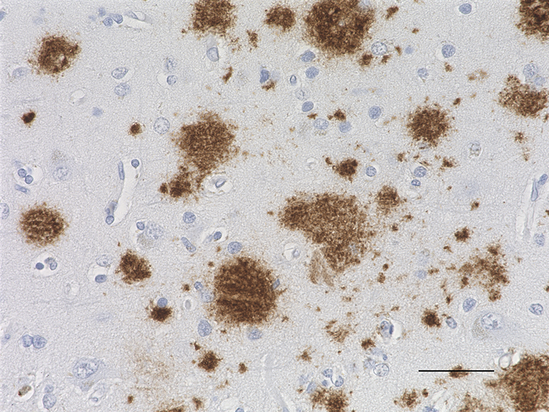
IHC staining of purified anti-Myelin Basic Protein antibody (clone SMI 99) on formalin-fixed paraffin-embedded human brain tissue. Following antigen retrieval using Retrieve-All Antigen Unmasking System 3: Acidic, 100X (Cat. No. 927601), the tissue was incubated with 0.25 µg/ml of the primary antibody overnight at 4°C, followed by incubation with Alexa Fluor® 647 goat anti-mouse IgG for one hour at room temperature. Nuclei were counterstained with DAPI, and the slides were mounted with ProLong™ Gold Antifade Mountant. The image was captured with a 20X objective. Scale Bar: 50 µm -

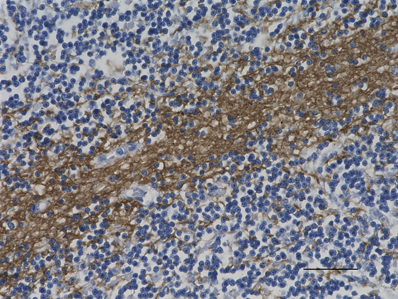
IHC staining of purified anti-Myelin Basic Protein antibody (clone SMI 99) on formalin-fixed paraffin-embedded human brain tissue. Following antigen retrieval using Retrieve-All Antigen Unmasking System 3: Acidic, 100X (Cat. No. 927601), the tissue was incubated with 0.25 µg/ml of the primary antibody overnight for 60 minutes at room temperature. BioLegend´s Ultra-Streptavidin (USA) HRP kit (Multi-Species, DAB, Cat. No. 929901) was used for detection followed by hematoxylin counterstaining, according to the protocol provided. The image was captured with a 10X objective. Scale bar: 50 µm -

IHC staining of purified anti-Myelin Basic Protein antibody (clone SMI 99) on formalin-fixed paraffin-embedded mouse brain tissue. Following antigen retrieval using Retrieve-All Antigen Unmasking System 3: Acidic, 100X (Cat. No. 927601), the tissue was incubated with 0.25 µg/ml of the primary antibody overnight at 4°C, followed by incubation with Alexa Fluor® 647 goat anti-mouse IgG for one hour at room temperature. Nuclei were counterstained with DAPI, and the slides were mounted with ProLong™ Gold Antifade Mountant. The image was captured with a 10X objective. Scale Bar: 50 µm -

IHC staining of purified anti-Myelin Basic Protein antibody (clone SMI 99) on formalin-fixed paraffin-embedded mouse brain tissue. Following antigen retrieval using Retrieve-All Antigen Unmasking System 3: Acidic, 100X (Cat. No. 927601), the tissue was incubated with 0.25 µg/ml of the primary antibody overnight for 60 minutes at room temperature. BioLegend´s Ultra-Streptavidin (USA) HRP kit (Multi-Species, DAB, Cat. No. 929901) was used for detection followed by hematoxylin counterstaining, according to the protocol provided. The image was captured with a 40X objective. Scale bar: 50 µm -

IHC staining of purified anti-Myelin Basic Protein antibody (clone SMI 99) on formalin-fixed paraffin-embedded rat brain tissue. Following antigen retrieval using Retrieve-All Antigen Unmasking System 3: Acidic, 100X (Cat. No. 927601), the tissue was incubated with 0.25 µg/ml of the primary antibody overnight at 4°C, followed by incubation with Alexa Fluor® 647 goat anti-mouse IgG for one hour at room temperature. Nuclei were counterstained with DAPI, and the slides were mounted with ProLong™ Gold Antifade Mountant. The image was captured with a 40X objective. Scale Bar: 50 µm -

Western blot of purified anti-Myelin Basic Protein antibody (clone SMI 99). Lane 1: Molecular weight marker; Lane 2: 10 µg of human brain lysate; Lane 3: 10 µg of mouse brain lysate; Lane 4: 10 µg of rat brain lysate. The blot was incubated with 1 µg/mL of the primary antibody overnight at 4°C, followed by incubation with HRP labeled goat anti-mouse IgG (Cat. No. 405306). Enhanced chemiluminescence was used as the detection system.
Myelin basic protein (MBP) is a protein involved in the myelination of nerves in the CNS. MBP functions to maintain the correct structure of myelin, through its interaction with the lipids in the myelin membrane. The myelin sheath is made up of MBP and lipids, and acts as an insulator to increase the velocity of axonal impulse conduction. MBP plays a central role in the demyelinating disease multiple sclerosis (MS). A hallmark of the disease is the loss of the myelin sheath surrounding nerves, thought to be induced by antibodies against MBP.
Product DetailsProduct Details
- Verified Reactivity
- Human, Mouse, Rat
- Antibody Type
- Monoclonal
- Host Species
- Mouse
- Formulation
-
Phosphate-buffered solution + 0.09% sodium azide.
Previous lots of this product may have been formulated with 0.1% or 0.05% NaN3 instead of 0.09% NaN3. For further information please contact BioLegend Technical Support or Customer Service. - Preparation
- The antibody was purified by affinity chromatography.
- Concentration
- 1 mg/ml
- Storage & Handling
- The antibody solution should be stored undiluted between 2°C and 8°C. Please note the storage condition for this antibody has been changed from -20°C to between 2°C and 8°C. You can also check your vial or your CoA to find the most accurate storage condition for this antibody.
- Application
-
IHC-P - Quality tested
WB - Verified
ICC - Reported in the literature, not verified in house - Recommended Usage
-
Each lot of this antibody is quality control tested by formalin-fixed paraffin-embedded immunohistochemical staining. For immunohistochemistry, a concentration range of 0.25 – 0.5 µg/ml is suggested. For Western blotting, the suggested use of this reagent is 0.2 – 1.0 µg per ml. It is recommended that the reagent be titrated for optimal performance for each application.
- Application Notes
-
This antibody reacts with the sequence Ala-Ser-Asp-Tyr-Lys-Ser in position 131-136 of the classic human myelin basic protein. The antibody detects myelin basic protein from most mammalian species. This antibody detects developing and adult myelin, developing oligodedrocytes and distinguishes oligodendrocytes from astrocytes, microglia, neurons, and other cells in brain sections. The combination of this antibody with clone SMI-91 and/or SMI-94 is useful for immunocytochemical studies on the progression of normal and pathologic myelination.
- Additional Product Notes
-
Myelin basic protein exists in multiple isoforms. By WB, clone SMI 99 detects mainly the 21 kD and 28 kD isoforms in the murine brain tissue, and the 21 kD isoform in the human brain tissue.
- Application References
-
- Fyffe-Maricich SL, et al. 2011. J. Neurosci. 31(3):843-50. (IHC, WB) PubMed
- Mendz GL, et al. 1985. Biochem. 228(1):61-8.
- Kerlero De Rosbo N, et al. 1984. Neurochem. Res. 9(10):1359-69.
- Carnegie PR, et al. 1983. J. Neuroimmunol. 5(2):125-34.
- Dowse CA, et al. 1983. J. Neuroimmunol. 5(2):135-44.
- Del Valle L,et al. Cancer Biol Ther. 9:286. (IHC) PubMed
- Kiryu-Seo S, et al. 2010. J. Neurosci. 30(19):6658-66. (ICC) PubMed
- Samanta J, et al. 2010. Stroke. 41(2):357-62. (IHC) PubMed
- Werner HB, et al. 2007. J. Neurosci. 27(29):7717-30. (WB, ICC) PubMed
- Product Citations
-
- RRID
-
AB_2734562 (BioLegend Cat. No. 808403)
AB_2564741 (BioLegend Cat. No. 808401)
AB_2564742 (BioLegend Cat. No. 808402)
Antigen Details
- Cell Type
- Oligodendrocytes
- Biology Area
- Cell Biology, Cell Structure, Neurodegeneration, Neuroinflammation, Neuroscience, Neuroscience Cell Markers
- Molecular Family
- Adhesion Molecules
- Gene ID
- 4155 View all products for this Gene ID 17196 View all products for this Gene ID 24547 View all products for this Gene ID
- UniProt
- View information about Myelin Basic Protein on UniProt.org

Related FAQs
Other Formats
View All Myelin Basic Protein Reagents Request Custom Conjugation| Description | Clone | Applications |
|---|---|---|
| Purified anti-Myelin Basic Protein | SMI 99 | IHC-P,WB,ICC |
| Alexa Fluor® 647 anti-Myelin Basic Protein | SMI 99 | IHC-P |
Customers Also Purchased
Compare Data Across All Formats
This data display is provided for general comparisons between formats.
Your actual data may vary due to variations in samples, target cells, instruments and their settings, staining conditions, and other factors.
If you need assistance with selecting the best format contact our expert technical support team.
-
Purified anti-Myelin Basic Protein

IHC staining of purified anti-Myelin Basic Protein antibody ... 
IHC staining of purified anti-Myelin Basic Protein antibody ... 
IHC staining of purified anti-Myelin Basic Protein antibody ... 
IHC staining of purified anti-Myelin Basic Protein antibody ... 
IHC staining of purified anti-Myelin Basic Protein antibody ... 
Western blot of purified anti-Myelin Basic Protein antibody ... -
Alexa Fluor® 647 anti-Myelin Basic Protein

IHC staining of Alexa Fluor® 647 anti-Myelin Basic Protein (... 
IHC staining of Alexa Fluor® 647 anti-Myelin Basic Protein (... 
IHC staining of Alexa Fluor® 647 anti-Myelin Basic Protein (...


Follow Us