 Login/Register


Using Flow Cytometry in Neuroscience Research
 |
Sample Preparation One of the initial hurdles in establishing flow cytometry applications in neuroscience was preparing suitable samples. When using whole brain tissue, generation of single cell suspensions is possible through a variety of methods. |
 |
 |
 |
| Mechanical Dissociation First, tissues need to be mechanically dissociated into small fragments using a razor blade prior to processing. |
Enzymatic Digestion Enzymes such as Trypsin, Accutase®, or Papain can be used to digest whole brain tissue samples. |
Density Gradient Percoll® or sucrose gradients help to remove myelin from the sample. For this, we use a 70/37/30% Percoll® gradient. |
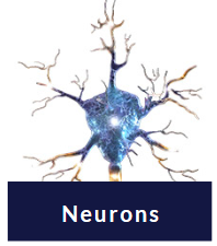
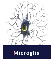
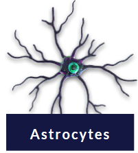
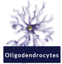
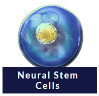
| Cell Markers Flow cytometry in neuroscience is useful when you need to study or isolate a homogenous population from a heterogeneous sample, such as primary cell suspensions or in vitro differentiation of neural stem cell cultures. Identification of cell type-specific markers allows for detection or isolation of these cells through magnetic cell separation or cell sorting. Cell type-specific markers have been identified for a variety of neural cell types including astrocytes, microglia, and oligodendrocytes. |
 |
 |
 |
| CD56 (NCAM) CD24 CD200 |
CD11b CD45 CX3CR1 P2RY12 TMEM119 |
GFAP GLAST |
 |
 |
||
| A2B5 PDGFRα NG2 |
|
Check out these references using flow cytometry to identify neural cell types: |
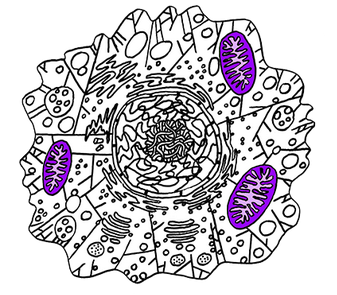 |
Organelle Markers & Function Altered organelle morphology and health is involved in numerous neurodegenerative diseases. For example, mitochondrial dysfunction and accumulation of reactive oxygen species is common in Alzheimer's and Parkinson's disease. When performing flow cytometry, in conjunction with cell type-specific markers, you can include a cell-permeant dye like MitoSpy™ as an indicator of cell health and mitochondrial function. Similarly, lysosomal proteins help clear aggregated proteins associated with disease. Antibodies against lysosome markers like LAMP-1 or LAMP-2 can be used in flow cytometry to compare lysosome function in normal and disease states. |
Find your antibodies for Neuroscience and Flow Cytometry at: biolegend.com/neuroscience |
| Contributed by Kelsey Swartz, PhD. |


Follow Us