 Login / Register


- Clone
- MECA-32 (See other available formats)
- Regulatory Status
- RUO
- Other Names
- Pan-endothelial Cell Antigen, MECA-32, Plasmalemma vesicle-associated protein
- Isotype
- Rat IgG2a, κ
- Ave. Rating
- Submit a Review
- Product Citations
- publications

-

bEND.3 cells stained with MECA-32 Alexa Fluor® 488
| Cat # | Size | Price | Quantity Check Availability | Save | ||
|---|---|---|---|---|---|---|
| 120501 | 50 µg | 57€ | ||||
| 120502 | 500 µg | 210€ | ||||
MECA-32 is a 50-55 kD homodimer glycoprotein primarily expressed on most endothelial cells in the embryonic and the adult mouse tissues, except in the brain and in skeletal and cardiac muscle, where it has more restricted distribution. In skeletal and cardiac muscle, the MECA-32 antigen is confined in small arterioles and venules. But in some conditions, such as inflammation or cardiac transplantation, MECA-32 expression can be induced on some negative-cardiac endothelial cells. In the brain, its expression occurs only in the circumventricular organs and the neurohemal tissues. During embryonic development, MECA-32 antigen is expressed on the brain vasculature up to day 16 of gestation, and disappears at day 17 and after.
Product DetailsProduct Details
- Verified Reactivity
- Mouse
- Antibody Type
- Monoclonal
- Host Species
- Rat
- Immunogen
- Mouse lymph node stromal cells
- Formulation
- Phosphate-buffered solution, pH 7.2, containing 0.09% sodium azide.
- Preparation
- The antibody was purified by affinity chromatography.
- Concentration
- 0.5 mg/ml
- Storage & Handling
- The antibody solution should be stored undiluted between 2°C and 8°C.
- Application
-
FC - Quality tested
ICC, IHC-F, IP, WB - Reported in the literature, not verified in house - Recommended Usage
-
Each lot of this antibody is quality control tested by immunofluorescent staining with flow cytometric analysis. For flow cytometric staining, the suggested use of this reagent is ≤ 0.25 µg per million cells in 100 µl volume. It is recommended that the reagent be titrated for optimal performance for each application.
- Application Notes
-
Additional reported applications (in the relevant formats) include: immunoprecipitation, Western blotting, immunocytochemistry, and immunohistochemical staining of acetone-fixed frozen sections2-5. Clone MECA-32 is not recommended for formalin-fixed paraffin sections.
- Application References
-
- Leppink DM, et al. 1989. Transplantation 48:874.
- Bergese SD, et al. 1994. Transplantation 57:711. (IHC)
- Tanneau GM, et al. 1999. J. Histochem. Cytochem. 47:1581. (IHC)
- Kopp HG, et al. 2005. Blood 106:505. (IHC)
- Hallmann R, et al. 1995. Dev. Dyn. 202:325. (IHC)
- Product Citations
-
- RRID
-
AB_493301 (BioLegend Cat. No. 120501)
AB_493302 (BioLegend Cat. No. 120502)
Antigen Details
- Structure
- 50-55 kD homodimer
- Distribution
-
Endothelial cell in the embryonic and the adult mice, except in the brain and in skeletal and cardiac muscle, where it has more restricted distribution
- Cell Type
- Embryonic Stem Cells, Endothelial cells
- Biology Area
- Cell Adhesion, Cell Biology, Immunology, Stem Cells
- Molecular Family
- Adhesion Molecules
- Antigen References
-
1. Leppnik DM, et al. 1989. Transplantation 48:874.
2. Hallmann R, et al. 1995. Dev. Dyn. 202:325.
3. Kruse A, et al. 1999. Bio. Reprod. 61:1393. - Gene ID
- 84094 View all products for this Gene ID
- UniProt
- View information about Panendothelial Cell Antigen on UniProt.org

Related FAQs
Other Formats
View All Panendothelial Cell Antigen Reagents Request Custom Conjugation| Description | Clone | Applications |
|---|---|---|
| Purified anti-mouse Panendothelial Cell Antigen | MECA-32 | FC,ICC,IHC-F,IP,WB |
| Biotin anti-mouse Panendothelial Cell Antigen | MECA-32 | FC |
| Alexa Fluor® 488 anti-mouse Panendothelial Cell Antigen | MECA-32 | FC |
| TotalSeq™-A0381 anti-mouse Panendothelial Cell Antigen | MECA-32 | PG |
| TotalSeq™-B0381 anti-mouse Panendothelial Cell Antigen Antibody | MECA-32 | PG |
Customers Also Purchased
Compare Data Across All Formats
This data display is provided for general comparisons between formats.
Your actual data may vary due to variations in samples, target cells, instruments and their settings, staining conditions, and other factors.
If you need assistance with selecting the best format contact our expert technical support team.
-
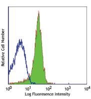
Purified anti-mouse Panendothelial Cell Antigen

bEND.3 cells stained with MECA-32 Alexa Fluor® 488 -
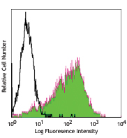
Biotin anti-mouse Panendothelial Cell Antigen

bEND.3 cells stained with MECA-32 biotin, followed by Sav-PE -
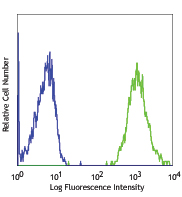
Alexa Fluor® 488 anti-mouse Panendothelial Cell Antigen
bEND.3 cells stained with MECA-32 Alexa Fluor® 488 -
TotalSeq™-A0381 anti-mouse Panendothelial Cell Antigen
-
TotalSeq™-B0381 anti-mouse Panendothelial Cell Antigen Antibody


Follow Us