 Login / Register


- Clone
- AA10 (See other available formats)
- Regulatory Status
- RUO
- Other Names
- β-3 Tubulin, Neuronal class III beta-tubulin, β3-tub
- Isotype
- Mouse IgG2a, κ
- Ave. Rating
- Submit a Review
- Product Citations
- publications

-

Day-three cultured postnatal C57BL/6 mouse brain cells were fixed with 1% paraformaldehyde (PFA) for ten minutes, permeabilized with 0.5 % Triton X-100 for ten minutes, and blocked with 5% FBS for 30 minutes. Then the cells were stained with 5 µg/mL of Alexa Fluor® 594 anti-Tubulin Beta 3 (TUBB3) (Clone AA10) (shown in red) in blocking buffer, overnight at 4°C, followed by staining with GFAP (shown in green) at room temperature for two hours. Nuclei were counterstained with DAPI (blue). The image was captured with 40X objective. -

C57BL/6 mouse frozen brain tissue was fixed with 4% paraformaldehyde (PFA) for ten minutes, permeabilized with 0.5 % Triton X-100 for ten minutes, and blocked with 5% FBS for 1 hour. Then the tissue was stained with 1.25 µg/mL of Alexa Fluor® 594 anti-Tubulin Beta 3 (TUBB3) (Clone AA10) (shown in red) and 5 µg/mL of Alexa Fluor® 488 anti-GFAP (Clone 2E1.E9) (shown in green) in blocking buffer, overnight at 4°C. Nuclei were counterstained with DAPI (blue). The image was captured with 10X objective. -

C57BL/6 mouse frozen brain tissue was fixed with 4% paraformaldehyde (PFA) for ten minutes, permeabilized with 0.5 % Triton X-100 for ten minutes, and blocked with 5% FBS for 30 minutes. Then the tissue was stained with 1.25 µg/mL of Alexa Fluor® 594 anti-Tubulin Beta 3 (TUBB3) (Clone AA10) (red) in blocking buffer, overnight at 4°C. Nuclei were counterstained with DAPI (blue). The image was captured with 10X objective. -

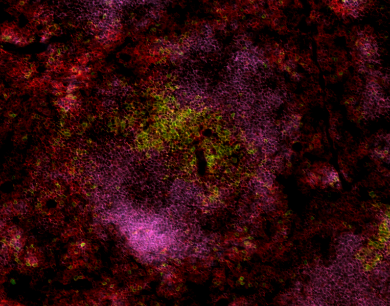
Human paraffin-embedded cerebellum tissue slices were prepared with a standard protocol of deparaffinization and rehydration. Antigen retrieval was done with Citrate Buffered 1X (1.0M, pH6.0) at 95°C for 40 minutes. Tissue was washed with PBS/0.05% Tween 20 twice for five minutes and blocked with 5% FBS and 0.2% gelatin for 30 minutes. Then, the tissue was stained with 10 µg/mL of Alexa Fluor® 647 anti-GFAP Antibody (Clone 2E1.E9, green) and Alexa Fluor® 594 anti-Tubulin Beta 3 (TUBB3) Antibody (Clone AA10, red) antibody overnight at 4°C. Nuclei were counterstained with DAPI (blue). The image was scanned with a 10X objective and stitched with MetaMorph® software. -

Human paraffin-embedded cerebellum tissue slices were prepared with a standard protocol of deparaffinization and rehydration. Antigen retrieval was done with Citrate Buffered 1X (1.0M, pH6.0) at 95°C for 40 minutes. Tissue was washed with PBS/0.05% Tween 20 twice for five minutes and blocked with 5% FBS and 0.2% gelatin for 30 minutes. Then, the tissue was stained with 10 µg/mL of Alexa Fluor® 647 anti-GFAP Antibody (Clone 2E1.E9, blue) and Alexa Fluor® 594 anti-Tubulin Beta 3 (TUBB3) Antibody (Clone AA10, red) antibody overnight at 4°C. Nuclei were counterstained with DAPI (green). The image was scanned with a 10X objective and stitched with MetaMorph® software. -
Formalin-fixed, 300 micron-thick mouse brain (cerebellum) section was blocked, permeabilized and stained overnight with Tubulin Beta 3 (TUBB3)(clone AA10) Alexa Fluor® 594 (green) and MAP2 (clone SMI 52) Alexa Fluor® 647 (magenta) both at 5 µg/mL, optically cleared, and analyzed on a confocal microscope. Watch the video.

| Cat # | Size | Price | Quantity Check Availability | Save | ||
|---|---|---|---|---|---|---|
| 657408 | 100 µg | 306€ | ||||
Tubulin is the main component of microtubules. In adults, tubulin beta 3 (TUBB3) is primarily expressed in neurons and is commonly used as a neuronal marker. It plays an important role in neuronal cell proliferation and differentiation. Mutations in this gene cause congenital fibrosis of the type 3 extraocular muscles. Tubulin beta 3 (TUBB3) is also found in a wide range of tumors. Studies indicate that it is a predictive and prognostic marker in various tumors.
Product DetailsProduct Details
- Verified Reactivity
- Mouse, Rat, Human
- Antibody Type
- Monoclonal
- Host Species
- Mouse
- Immunogen
- Fusion protein
- Formulation
- Phosphate-buffered solution, pH 7.2, containing 0.09% sodium azide.
- Preparation
- The antibody was purified by affinity chromatography and conjugated with Alexa Fluor® 594 under optimal conditions.
- Concentration
- 0.5 mg/mL
- Storage & Handling
- The antibody solution should be stored undiluted between 2°C and 8°C, and protected from prolonged exposure to light. Do not freeze.
- Application
-
IHC-F - Quality tested
ICC, IHC-P, 3D IHC - Verified - Recommended Usage
-
Each lot of this antibody is quality control tested by immunohistochemical staining on frozen tissue sections. For immunohistochemical staining on frozen tissue sections, the suggested use is 0.6 - 5 µg/mL. For immunocytochemistry microscopy, a concentration range of 2.5 - 5.0 μg/mL is recommended. For immunohistochemical staining on formalin-fixed paraffin-embedded tissue sections, the suggested use of this reagent is 5.0 - 10 µg per mL. It is recommended that the reagent be titrated for optimal performance for each application.
* Alexa Fluor® 594 has an excitation maximum of 590 nm, and a maximum emission of 617 nm.
Alexa Fluor® and Pacific Blue™ are trademarks of Life Technologies Corporation.
View full statement regarding label licenses - Application Notes
-
Additional reported application (for relevant formats) include: spatial biology (IBEX)1,2.
- Application References
- Product Citations
-
- RRID
-
AB_2565285 (BioLegend Cat. No. 657408)
Antigen Details
- Structure
- 450 amino acids with predicted molecular weight of 50 kD
- Distribution
-
Cytosol
- Function
- Plays important roles in neuronal cell proliferation and differentiation
- Interaction
- Alpha tubulin, kinesin and dynein
- Cell Type
- Mature Neurons
- Biology Area
- Cell Biology, Cell Cycle/DNA Replication, Cell Motility/Cytoskeleton/Structure, Immunology, Neuroscience, Neuroscience Cell Markers
- Molecular Family
- Microtubules
- Antigen References
-
1. Katsetos CD, et al. 2003. J. Child Neurol. 18:851.
2. Mobarakeh ZT, et al. 2012. Cell Biol. Int. Rep. (2010) 19:e00015.
3. Locher H, et al. 2013. Differentiation. 85:173.
4. Karki R, et al. 2013. Expert Opin. Ther. Targets. 17:461.
5. Mariani M, et al. 2011. Curr. Mol. Med. 11:726.
6. Koh Y, et al. 2009. Ann. Oncol. 20:1414. - Gene ID
- 10381 View all products for this Gene ID
- UniProt
- View information about Tubulin beta-3 on UniProt.org


Related FAQs
Other Formats
View All Tubulin β-3 (TUBB3) Reagents Request Custom Conjugation| Description | Clone | Applications |
|---|---|---|
| Purified anti-Tubulin Beta 3 (TUBB3) | AA10 | WB,ICC |
| Alexa Fluor® 488 anti-Tubulin Beta 3 (TUBB3) | AA10 | ICFC,IHC-F,3D IHC |
| Alexa Fluor® 647 anti-Tubulin Beta 3 (TUBB3) | AA10 | ICFC,IHC-F,3D IHC,SB |
| Alexa Fluor® 594 anti-Tubulin Beta 3 (TUBB3) | AA10 | IHC-F,ICC,IHC-P,3D IHC |
| Direct-Blot™ HRP anti-Tubulin Beta 3 (TUBB3) | AA10 | WB |
| Brilliant Violet 421™ anti-Tubulin Beta 3 (TUBB3) | AA10 | IHC-F,ICC,SB |
Customers Also Purchased
Compare Data Across All Formats
This data display is provided for general comparisons between formats.
Your actual data may vary due to variations in samples, target cells, instruments and their settings, staining conditions, and other factors.
If you need assistance with selecting the best format contact our expert technical support team.
-
Purified anti-Tubulin Beta 3 (TUBB3)


Mouse brain (lane 1), mouse liver (lane 2), N-tera2 (lane 3)... 
Whole cell lysates (15 µg protein) from HEL (negative contro... 
NTERA‐2 cells were fixed with 4% paraformaldehyde (PFA) for ... -
Alexa Fluor® 488 anti-Tubulin Beta 3 (TUBB3)

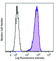
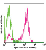
Human lung adenocarcinoma cell line A549 was treated with Bi... 
C57BL/6 mouse frozen brain tissue was fixed with 4% paraform... 
Dissected C57/B6 mouse small intestine was immersed in 4% p... 
Formalin-fixed, 300 micron-thick mouse kidney section was bl... -
Alexa Fluor® 647 anti-Tubulin Beta 3 (TUBB3)

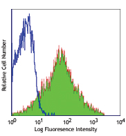
Human lung adenocarcinoma cell line A549 was treated with Fi... 
C57BL/6 mouse frozen brain tissue was fixed with 4% paraform... Formalin-fixed, 300 micron-thick mouse large intestine secti... -
Alexa Fluor® 594 anti-Tubulin Beta 3 (TUBB3)

Day-three cultured postnatal C57BL/6 mouse brain cells were ... 
C57BL/6 mouse frozen brain tissue was fixed with 4% paraform... 
C57BL/6 mouse frozen brain tissue was fixed with 4% paraform... 
Human paraffin-embedded cerebellum tissue slices were prepar... 
Human paraffin-embedded cerebellum tissue slices were prepar... Formalin-fixed, 300 micron-thick mouse brain (cerebellum) se... -
Direct-Blot™ HRP anti-Tubulin Beta 3 (TUBB3)

10 µg of total protein extract from HeLa cells was resolved ... -
Brilliant Violet 421™ anti-Tubulin Beta 3 (TUBB3)

Human lung adenocarcinoma cell line A549 was fixed with 1% p... 
C57BL/6 mouse frozen brain (cerebellum) tissue was fixed wit...


Follow Us