 Login / Register


- Clone
- N308/48 (See other available formats)
- Regulatory Status
- RUO
- Other Names
- GluN1/NR1 glutamate receptor, Glutamate receptor ionotropic, NMDA 1, NMD-R1, neurotransmitter receptor, NMDAR1 receptor C1 cassette, N-methyl-D-aspartate glutamate receptor, glutamate receptor subunit zeta-1, N-methyl-D-aspartate receptor subunit NR1
- Previously
-
Covance Catalog# MMS-5145
- Isotype
- Mouse IgG1, κ
- Ave. Rating
- Submit a Review
- Product Citations
- publications

-

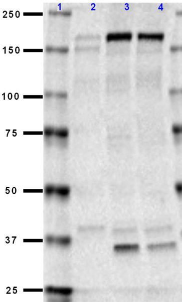
Western blot of purified anti-NMDAR1 antibody (clone N308/48). Lane 1: Molecular weight marker; Lane 2: 20 µg of human brain membrane lysate; Lane 3: 20 µg of mouse brain membrane lysate; Lane 4: 20 µg of rat brain membrane lysate. The blot was incubated with 10 µg/mL of the primary antibody overnight at 4°C. Enhanced chemiluminescence (Cat. No. 426303) was used as the detection system. The smaller band at ~75 kD is an MMP-7 cleaved fragment of NMDAR1. -

IHC staining of purified anti-NMDAR1 antibody (clone N308/48) on formalin-fixed paraffin-embedded mouse brain tissue. Following antigen retrieval using Sodium Citrate H.I.E.R., the tissue was incubated with 10 µg/mL of the primary antibody overnight at 4°C. BioLegend´s Ultra-Streptavidin (USA) HRP kit (Multi-Species, DAB, Cat. No. 929901) was used for detection followed by hematoxylin counterstaining, according to the protocol provided. The image was captured with a 40X objective. Scale bar: 50 µm -

IHC staining of purified anti-NMDAR1 antibody (clone N308/48) on formalin-fixed paraffin-embedded rat brain tissue. Following antigen retrieval using Sodium Citrate H.I.E.R., the tissue was incubated with 10 µg/ml of the primary antibody overnight at 4°C. BioLegend´s Ultra-Streptavidin (USA) HRP kit (Multi-Species, DAB, Cat. No. 929901) was used for detection followed by hematoxylin counterstaining, according to the protocol provided. The image was captured with a 40X objective. Scale bar: 50 µm
| Cat # | Size | Price | Quantity Check Availability | Save | ||
|---|---|---|---|---|---|---|
| 818602 | 25 µL | 67€ | ||||
| 818601 | 100 µL | 132€ | ||||
Glutamate activated Ion channels that are sensitive to N-methyl-D-aspartate (NMDA) are designated NMDA receptors (NMDAR). The NMDA receptor plays an essential role in the induction of LTP in the CA1 and dentate areas of the hippocampus as the specific NMDA antagonist, APV blocks LTP in these areas. This receptor has also been linked to neuronal development and it has been implicated in several disorders of the central nervous system including epilepsy and ischemic neuronal cell death. The NR1 protein can form NMDA activated channels when expressed in Xenopus oocytes but the currents in such channels are much smaller than those seen in situ. Channels with more physiological characteristics are produced when the NR1 subunit is combined with one or more of the NMDAR2 (NR2 A-D) subunits.
Product DetailsProduct Details
- Verified Reactivity
- Human, Rat, Mouse
- Antibody Type
- Monoclonal
- Host Species
- Mouse
- Immunogen
- This monoclonal antibody was raised against a fusion protein corresponding to amino acids 42-361 (extracellular, N-terminus) of rat GluN1/NR1 glutamate receptor protein.
- Formulation
- Phosphate-buffered solution.
- Preparation
- The antibody was purified by affinity chromatography.
- Concentration
- 1 mg/mL
- Storage & Handling
- The antibody solution should be stored undiluted between 2°C and 8°C. Please note the storage condition for this antibody has been changed from -20°C to between 2°C and 8°C. You can also check your vial or your CoA to find the most accurate storage condition for this antibody.
- Application
-
WB - Quality tested
IHC-P - Verified - Recommended Usage
-
Each lot of this antibody is quality control tested by Western blotting. For Western blotting, the suggested use of this reagent is 10 µg/mL. For immunohistochemistry on paraffin-embedded tissue sections, a concentration of 10 µg/mL is suggested. It is recommended that the reagent be titrated for optimal performance for each application.
- Product Citations
-
- RRID
-
AB_2734577 (BioLegend Cat. No. 818602)
AB_2564822 (BioLegend Cat. No. 818601)
Antigen Details
- Structure
- Expected MW: 105 kD
- Cell Type
- Glutamatergic Neurons
- Biology Area
- Cell Biology, Neuroscience, Neuroscience Cell Markers, Synaptic Biology
- Molecular Family
- Postsynaptic proteins
- Gene ID
- 24408 View all products for this Gene ID
- UniProt
- View information about NMDAR1 on UniProt.org


Related FAQs
Other Formats
View All NMDAR1 Reagents Request Custom Conjugation| Description | Clone | Applications |
|---|---|---|
| Purified anti-NMDAR1 | N308/48 | WB,IHC-P |
| Biotin anti-NMDAR1 | N308/48 | IHC-P |
Customers Also Purchased
Compare Data Across All Formats
This data display is provided for general comparisons between formats.
Your actual data may vary due to variations in samples, target cells, instruments and their settings, staining conditions, and other factors.
If you need assistance with selecting the best format contact our expert technical support team.
-
Purified anti-NMDAR1

Western blot of purified anti-NMDAR1 antibody (clone N308/48... 
IHC staining of purified anti-NMDAR1 antibody (clone N308/48... 
IHC staining of purified anti-NMDAR1 antibody (clone N308/48... -
Biotin anti-NMDAR1

IHC staining of Biotin anti-NMDAR1 antibody (clone N308/48) ... 
IHC staining of Biotin anti-NMDAR1 antibody (clone N308/48) ... 
IHC staining of Biotin anti-NMDAR1 antibody (clone N308/48) ...


Follow Us