The neuron is a critical functional unit of the nervous system, which relays electrical and chemical signals to other neurons at junctions known as the neural synapse. The differentiated mammalian central nervous system is estimated to contain at least 100 billion neurons, which communicate with each other through numerous synaptic connections. The neural synapse most often occurs between the axon of one neuron and the dendrites of another and is composed of hundreds of proteins that function together to coordinate the exquisitely tuned signals that are the physical basis for higher nervous system functions, such as cognition, memory, and movement. BioLegend provides an extensive selection of antibodies and reagents for the analysis of synaptic function.


We've permanently discounted over 70 synaptic biology antibody products by up to 40%View the products...

image

Many distinct types of electrical synapses exist that relay hundreds of known chemical signals, termed neurotransmitters, from one neuron to another, or form a direct contact. Synapses can weaken or strengthen in response to a number of factors, a phenomenon which is termed neural plasticity. Neurodegenerative diseases, such as Alzheimer's and Parkinson's, display a loss of synaptic function and a subsequent degradation of these structures. This degeneration occurs more prominently at particular locations in the central nervous system, such as the substantia nigra for Parkinson's disease, which is highly enriched in synapses that transmit the neurotransmitter dopamine. With the support of BioLegend reagents, investigations into the mechanisms that lead to dysfunction and synaptic loss, may provide new avenues for preventative and therapeutic intervention.

Featuring

Purified anti-MAGUK (pan reactive)

/Files/Images/media_assets/products/product_images/K28slash86_Purified_MAGUK_Antibody_1_020718.png

Membrane-associated guanylate kinases (MAGUK) are a superfamily of scaffolding proteins defined by the presence of a guanylate kinase (GUK) domain. MAGUKs, such as PSD-95, SAP97, PSD-83, CASK, and MAGIs are involved in the synaptic development and function where they are important for the spatial organization of both pre- and postsynaptic proteins, including glutamate and other receptors.

See More

Purified anti-Ankyrin-B

/Files/Images/media_assets/products/product_images/N105slash13__PURE_Ankyrin-B_Antibody_1_021918.png

Ankyrins are adaptor proteins that mediate the attachment of membrane proteins to the cytoskeleton. Ankyrin-B (Ankyrin-2) is highly expressed in cardiac tissue where it has an essential role in stabilizing ion transporters and channels in cardiomyocytes in the membrane. Mutations in ankyrin-B are associated with cardiac arrhythmia and ankyrin-B may be involved in human heart failure.

See More

Purified anti-PSD95

/Files/Images/media_assets/products/product_images/K28slash43_PURE_PSD95_Antibody_WB_051418.png

PSD95, also known as SAP-90, is a member of the MAGUK family, encoded by the the DLG4 gene.  At postsynaptic sites, it mediates the scaffolding of NMDA receptors, AMPA receptors, potassium channels, and other associated signaling protein clusters. It plays an important role in synaptic plasticity and the stabilization of synaptic changes during long-term potentiation.

See More

Purified anti-Neuroligin-1

/Files/Images/media_assets/products/product_images/N97Aslash31_Purified_Neuroligin-1_Antibody_1_032218.png

Neuroligin is a postsynaptic membrane protein that binds to Neurexin, helping to form and maintain neuronal synapses.  Within the postsynaptic neuron, it binds to proteins such as PSD-95, which recruits receptors and channels to the synapse.

See More

Purified anti-Synaptotagmin-12

/Files/Images/media_assets/products/product_images/N2777_PURE_Synaptotagmin12_Antibody_HR_11_090517.jpg

Synaptotagmin 12 (Syt12) is regulated by protein kinase A (PKA) and is unique from other synaptotagmins in that it does not bind to Ca2+. Syt12 interaction with Syt1 prevents SNARE complex binding. Expression of Syt12 induces spontaneous neurotransmitter release, providing a mechanism for spontaneous synaptic-vesicle exocytosis that is Ca2+-independent.

See More

A complex array of proteins mediate the interconnection between the presynaptic membrane and the postsynaptic membrane. Adhesion molecules also contribute to a variety of processes, including the creation of synapses, mediating and modifying communication through the synapse, and modulating synaptic plasticity.
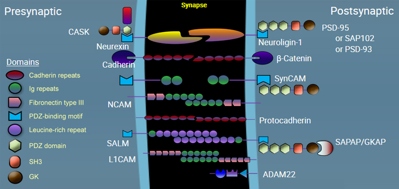


Neurexin

Neurexin is a presynaptic membrane adhesion molecule on neurons, important in the formation and specificity of synapses. It binds to neuroligin in the neuronal synapse. Within the presynaptic neuron, it associates with vesicular trafficking proteins including CASK and Mint.


Neuroligin-1

Neuroligin is a postsynaptic membrane protein that binds to Neurexin, helping to form and maintain neuronal synapses. Within the postsynaptic neuron, it binds to proteins such as PSD-95, which recruits receptors and channels to the synapse.
 

View Neuroligin-1 products


Cadherin

CD325 (N-cadherin) is important for neuronal migration during development, neurite outgrowth and target finding during differentiation, initial formation of synapses, and synaptic plasticity in mature neurons. CD325 (N-cadherin) is a 130 kD single pass transmembrane protein with an extracellular region consists of five cadherin domains. N-cadherin is involved in organogenesis and in the maintenance of organ architecture by contributing to the sorting of heterogeneous cell types and in the cell adhesion needed to form tissues. N-cadherin is expressed by stem cells, myeloblasts, endothelial cells, fibroblasts, and also is expressed in neural and muscle tissues along with some types of carcinoma cells. CD325 associates with the cytoskeleton through catenin proteins.

View Cadherin products


SynCAM

SynCAM molecules mediate homotypic adhesion in the neural synapse. These proteins are also known as Necl1-4 or cell adhesion molecule 1-4.

View SynCAM products


NCAM

CD56 is a single transmembrane glycoprotein also known as NCAM (neural cell adhesion molecule), Leu-19, or NKH1. It is a member of the Ig superfamily. The 140 kD isoform is expressed on NK and NKT cells. CD56 is also expressed in the brain (cerebellum and cortex) and at neuromuscular junctions. Certain large granular lymphocyte (LGL) leukemias, small-cell lung carcinomas, neuronal derived tumors, myelomas, and myeloid leukemias also express CD56. CD56 plays a role in homophilic and heterophilic adhesion via binding to itself or heparan sulfate.

View NCAM products


Protocadherin

Protocadherins are a group of self adhering molecules belonging to the cadherin superfamily of proteins. They are predominantly expressed in neurons in widely diverse array. Unlike cadherin proteins, protocadherins do not bind to intracellular catenin molecules.

View Protocadherin products


SALM

SALM, also known as LRFN, is homotypic adhesion molecule that promotes neurite outgrowth in hippocampal neurons, regulates excitatory synapses, and induces the clustering of excitatory postsynaptic proteins, including DLG4, DLGAP1, GRIA1 (AMPA receptor subunit) and GRIN1 (NMDA receptor subunit).


L1CAM

L1CAM, also known as CD171, is a neuronal cell adhesion molecule involved in axon guidance and cell migration with a strong implication in treatment-resistant cancers. This cell adhesion molecule plays an important role in nervous system development, including neuronal migration, and differentiation. Mutations in the gene cause three X-linked neurological syndromes known by the acronym CRASH (corpus callosum hypoplasia, retardation, aphasia, spastic paraplegia and hydrocephalus). CD171 has been shown to function as a cell adhesion molecule mediating homotypic and heterotypic cell-cell interactions in neuronal myelination, neurite outgrowth and regeneration.

View L1CAM products


ADAM22

ADAM22 is a member of the ADAM (a disintegrin and metalloprotease domain) family, implicated in a variety of biological processes involving cell-cell and cell-matrix interactions, including fertilization, muscle development, and neurogenesis. Unlike other members of the ADAM protein family, the protein encoded by this gene lacks metalloprotease activity since it has no zinc-binding motif. The protein is highly expressed in the brain and may function as an integrin ligand in the brain. In mice, it has been shown to be essential for correct myelination in the peripheral nervous system.

View ADAM22 products


CASK

Calcium/Calmodulin-Dependent Serine Protein Kinase (CASK) is a multidomain scaffolding protein with a role in synaptic transmembrane protein anchoring and ion channel trafficking. It contributes to neural development and regulation of gene expression via interaction with the transcription factor TBR1. It binds to cell-surface proteins, including amyloid precursor protein, neurexins and syndecans.

View CASK products


PSD-95

PSD95, also known as SAP-90, is a member of the MAGUK family, encoded by the the DLG4 gene. At postsynaptic sites, it mediates the scaffolding of NMDA receptors, AMPA receptors, potassium channels, and other associated signaling protein clusters. It plays an important role in synaptic plasticity and the stabilization of synaptic changes during long-term potentiation.

View PSD-95 products


SAP-102

SAP-102, also known as DLG3 or neuroendocrine-DLG, is a member of the MAGUK (membrane-associated guanylate kinase) superfamily. It interacts with a number of adhesion molecules as well as glutamate receptors on the postsynaptic membrane, mediating scaffolding of synaptic signaling.

View SAP-102 products


PSD-93

PSD-93, also known as DLG2 or Chapsyn-110, is a member of the MAGUK family important in the scaffolding of receptors with adhesion molecules on the postsynaptic membrane. It regulates surface expression of receptors by directly interacting with the cytoplasmic tail of NMDA receptor subunits and inward rectifying potassium channels, and is involved in the regulation of synaptic stability at synapses.

View PSD-93 products


SAPAP

SAPAP is also known as DAP-1 or GKAP, encoded by the DLGAP1 gene. It is enriched in the postsynaptic membrane and is responsible for connecting PSD-95 and SHANK, which serves as an adapter protein that interconnects receptors of the postsynaptic membrane including NMDA-type and metabotropic glutamate receptors, and the actin-based cytoskeleton.

View SAPAP products


β-Catenin

β-catenin is part of a complex of proteins that constitute adherens junctions (AJs). AJs are necessary for the creation and maintenance of epithelial cell layers by regulating cell growth and adhesion between cells. The encoded protein also anchors the actin cytoskeleton and may be responsible for transmitting the contact inhibition signal that causes cells to stop dividing once the epithelial sheet is complete. β-catenin also plays a key role in Wnt signaling pathways and thus is involved in neural differentiation, synaptic plasticity, neurodegenerative disease, and prevention of apoptosis. Finally, this protein binds to the product of the adenomatous polyposis coli (APC) gene, which is mutated in the adenomatous polyposis of the colon. Mutations in this gene are a cause of colorectal cancer (CRC), pilomatrixoma (PTR), medulloblastoma (MDB), and ovarian cancer.

View β-Catenin products

There are two types of neurotransmitter receptors in the synapse: ligand-gated ion channels (ionotropic receptors) and G-protein coupled receptors (metabotropic receptors). Ionotropic receptors are composed of five transmembrane protein subunits that form a single pore that spans the membrane. Activation of the receptor by neurotransmitters causes the pore to open and allows the direct passage of ions such as Ca2+, Na+, K+, or Cl- through the membrane. These receptors can be excited or inhibited depending on the type of ligand they bind to. Ligands such as glutamate and aspartate are excitatory, whereas GABA and glycine are inhibitory.


Metabotropic receptors are seven-transmembrane domain receptors that unlike ionotropic receptors, do not form a membrane-spanning pore. Instead, when a neurotransmitter binds to the extracellular ligand-binding site, these receptors couple with and activate an intermediate molecule within the postsynaptic cell, called a G-protein. Activation of G proteins initiates a signal transduction cascade that involves second messengers to open or close ion channels located at other places on the cell membrane. Metabotropic receptors are associated with a slow action and more prolonged stimulus since their action is not as direct compared to ionotropic receptors.


Adrenergic Receptors

These belong to a class of G protein-coupled receptors that bind to norepinephrine (noradrenaline) and epinephrine (adrenaline). Adrenergic Receptors are composed of two main types, a and b, and further subdivided into several subtypes: α1A, α1B, α1C, α1D, α2A, α2B, α2C, α2D, β1, β2, β3. Combination of the different subtypes of the receptor generally determines whether the response it transmits will be stimulatory or inhibitory.


Dopaminergic Receptors

These are a class of G protein-coupled receptors that bind to the neurotransmitter dopamine. These receptors regulate a range of neurological processes including voluntary movement, memory, reward and hormonal regulation. There are five known subtypes of the dopamine receptor: D1, D2, D3, D4, D5. After dopamine is released into the synapse, it is re-accumulated into cells and stored in vesicles for later release by an integral membrane protein called Dopamine Transporter (DAT).

View Dopamine products


GABAergic Receptors

These receptors respond to Gamma-Aminobutyric Acid (GABA), which is the major inhibitory neurotransmitter in the mammalian brain. There are two classes of GABA receptors: GABAA which are ligand-gated ion channels, and GABAB, that are G protein-coupled receptors. Upon activation, the GABAA receptor selectively conducts Cl− through its pore, resulting in hyperpolarization of the neuron. The GABAA receptor is generally pentameric and constitutes of several subunits which determines the receptor's conductance, affinity to agonists and other properties. 

In humans, the subunits are:

  • α subunits: GABRA1, GABRA2, GABRA3, GABRA4, GABRA5, GABRA6
  • β subunits: GABRB1, GABRB2, GABRB3
  • γ subunits: GABRG1, GABRG2, GABRG3
  • Other subunits: δ (GABRD), ε (GABRE), π (GABRP), and θ (GABRQ).


The GABAB receptors are usually considered as inhibitory receptors and they are linked to K+ channels in the cell through G-proteins. There are two subtypes of this receptor, GABAB1 (also known as GABA(B) receptor 1) and GABAB2 (GABA(B) receptor 2). 

View GABA receptor products


Glutaminergic Receptors

These receptors bind to glutamate which is the major excitatory neurotransmitter in the human brain. They are important in memory formation, learning and modulation of synaptic plasticity. Glutamate receptors can be broadly divided into two groups: ionotropic glutamate receptors (iGluRs) and metabotropic glutamate receptors (mGluRs). iGluRs include NMDA receptors, Kainate receptors and AMPA receptors, mGluRs include mGluR1, mGluR2, mGluR3, mGluR4, mGluR5, mGluR6, mGluR7.

View NMDA products
View GluR products


Histaminergic Receptors

These receptors belong to the class of G protein–coupled receptors. They primarily bind to histamine. There are four types of histamine receptors: H1, H2, H3 and H4.


Cholinergic Receptors

These receptors primarily respond to the neurotransmitter acetylcholine so they are also known as acetylcholine receptors. Besides acetylcholine, these receptors can also bind to nicotine and muscarine so they are also referred to as nicotinic acetylcholine receptors (nAChR) and muscarinic acetylcholine receptors mAChR respectively. The nAChRs are ligand-gated ion channels, whereas the mAChRs belong to the family of G-protein-coupled receptors.


Opiod Receptors

These belong to the family of G protein-coupled receptors and they respond to opiod. Common examples of endogenous opioids are endorphins, endomorphins, etc. There are four major subtypes of this receptor: µ, δ1/2, κ and nociceptin receptor. 

View Opioid receptor products


Serotonergic Receptors

These receptors are also known as 5-hydroxytryptamine receptors or 5-HT receptors and are activated by the neurotransmitter serotonin, which is their natural ligand. These receptors include both G protein-coupled receptors and ligand-gated ion channels and they mediate both excitatory and inhibitory neurotransmission. There are 7 known types of 5-HT receptors (5-HT1-7) and these are further subdivided into 14 subtypes.


Glycinergic Receptors

These receptors are ionotropic receptors that bind to glycine. They primarily transmit Cl- ions through the membrane pore which mediates inhibitory neurotransmission.


In a chemical synapse, signals are transmitted via release of neurotransmitters that are packed in synaptic vesicles in the presynaptic neuron, into the synaptic cleft. This release of neurotransmitters is regulated by a voltage-dependent calcium channel. The release of neurotransmitters at the synapse are constantly recycled through repeating steps of trafficking, exocytosis and endocytosis as shown in the diagram below. Furthermore, this process is coordinated through the action of about 50-100 different proteins.


1. Storage of synaptic vesicle in a reserve pool

Synaptic vesicles are contained in several different reserves that are mobilized following an action potential to replenish vesicles that have undergone fusion. Synapsins are a family of proteins that regulate the migration of the synaptic vesicles from these reserve pools to the membrane for neurotransmitter release.


View Synapsin products


2. Trafficking to the synapse

Synaptic vesicle trafficking is a multistep process involving several proteins and lipids. Synapsin, members of the kinesin motor family, and ATP produced by the mitochondria, are some of the factors implicated in the trafficking of synaptic vesicles to the membrane.


View Synapsin products


3. Transmitter loading

Vesicles are loaded with neurotransmitters when they are at the synaptic site. This is an active process involving transporters and a vacuolar-type proton pump ATPase that provides a pH and electrochemical gradient. These transporters are selective for different classes of transmitters.


View transporter protein products


4. Docking

Docking is the first step in the secretory pathway before vesicles fuse with the pre-synaptic membrane to release their contents. Once at the active zone, vesicle proteins, as well as, plasma membrane and cytoplasmic proteins, initiate the process of docking.


5. Priming

After initial docking, vesicles need to be ‘primed’ because they are not fusion competent. This priming step is thought to involve the formation of partially assembled SNARE (Soluble NSF Attachment Protein Receptor) complexes.


6. Fusion

Following priming, vesicles fuse rapidly in response to calcium influx. This membrane fusion event is thought to be mediated directly by the formation of the SNARE (Soluble NSF Attachment Protein Receptor) complex. This complex consists of the vesicle-associated Synaptobrevin, and the plasma membrane-associated Syntaxin and SNAP25, that bridges the synaptic vesicle and plasma membrane together. The calcium sensor which positively regulates SNARE-dependent fusion of vesicles with membranes is Synaptotagmin.


View SynaptotagminDocSynaptobrevinSyntaxin, or SNAP25 products


7. Exocytosis

Membrane fusion connects the lumen of the vesicle with the synaptic cleft forming a pore, similar to a gap junction, leading to exocytosis and neurotransmitter release. There are two leading mechanisms of action by which this happens: full collapse fusion and the "kiss-and-run" method. During full collapse fusion, after release of the neurotransmitter, the pore dilates fully so that the vesicle collapses completely into the synaptic membrane, whereas for kiss-and-run fusion the vesicle transiently fuses with the plasma membrane and the pore closes rapidly by pinching off the membrane. Proteins potentially involved in forming the fusion pore include synaptophysin on the vesicle membrane and physophilin on the pre-synaptic membrane. 


View Synaptophysin Products
View Dynamin Products


8. Endocytosis and endosomal fusion

After fusion, synaptic vesicle membrane is recovered via clathrin-mediated endocytosis that is based on a membrane budding process and requires the formation of coated pits and coated vesicles. Several regulatory proteins such as endophilin, synaptojanin, and dynamin play a role to coordinate this process. As a final step in the recycling process, it has been proposed that clathrin-coated vesicles de-coat and subsequently fuse with early endosomes. New synaptic vesicles then regenerate by budding from the endosome. 


View Clathrin Products
View Dynamin Products


ProductsHere

Login/Register
Remember me
Forgot your password? Reset Password
Request an Account