 Login / Register


- Clone
- SMI 25 (See other available formats)
- Regulatory Status
- RUO
- Other Names
- Glial fibrillary acidic protein
- Isotype
- Mouse IgG2b, κ
- Ave. Rating
- Submit a Review
- Product Citations
- publications

-

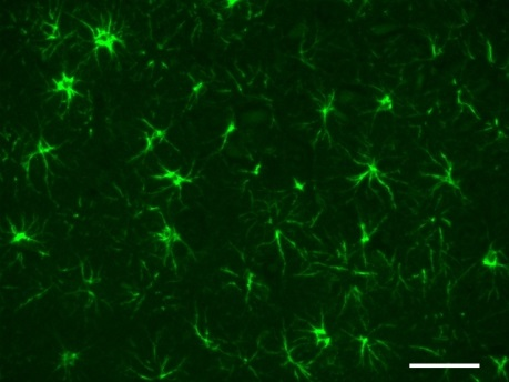
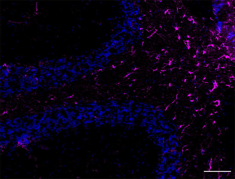
IHC staining of Alexa Fluor® 647 anti-GFAP antibody (clone SMI 25) on formalin-fixed paraffin-embedded rat brain tissue. Following antigen retrieval using Retrieve‐All Antigen Unmasking System 3 (Cat. No. 927801), the tissue was incubated with the primary antibody at 10 µg/ml overnight at 4°C. Nuclei were counterstained with DAPI. The image was captured with a 40X objective. Scale bar: 50 µm -

IHC staining of Alexa Fluor® 647 anti-GFAP antibody (clone SMI 25) on formalin-fixed paraffin-embedded mouse brain tissue. Following antigen retrieval using Retrieve‐All Antigen Unmasking System 3 (Cat. No. 927801), the tissue was incubated with the primary antibody at 5 µg/ml overnight at 4°C. Nuclei were counterstained with DAPI. The image was captured with a 40X objective. Scale bar: 50 µm -

IHC staining of Alexa Fluor® 647 anti-GFAP antibody (clone SMI 25) on formalin-fixed paraffin-embedded human brain tissue. Following antigen retrieval using Retrieve‐All Antigen Unmasking System 3 (Cat. No. 927801), the tissue was incubated with the primary antibody at 10 µg/ml overnight at 4°C. Nuclei were counterstained with DAPI. The image was captured with a 40X objective. Scale bar: 50 µm
| Cat # | Size | Price | Quantity Check Availability | Save | ||
|---|---|---|---|---|---|---|
| 837511 | 25 µg | 104€ | ||||
| 837512 | 100 µg | 259€ | ||||
Glial fibrillary acidic protein is an intermediate filament (IF) protein that is expressed by numerous cell types of the central nervous system (CNS) including astrocytes and ependymal cells. GFAP has also been found to be expressed in glomeruli and peritubular fibroblasts, Leydig cells of the testis, keratinocytes, osteocytes and chondrocytes and stellate cells of the pancreas and liver. GFAP is a type III IF protein that is closely related to its non-epithelial family members, vimentin, desmin, and peripherin, which are all involved in the structure and function of the cell’s cytoskeleton. GFAP is thought to help to maintain astrocyte mechanical strength, as well as the shape of cells.
Type III intermediate filaments are highly conserved and contain three domains, named the head, rod and tail domains. This rod domain coils around that of another filament to form a dimer, with the N-terminal and C-terminal of each filament aligned. Type III filaments such as GFAP are capable of forming both homodimers and heterodimers; GFAP can polymerize with other type III proteins or with neurofilament protein (NF-L). Interestingly, GFAP and other type III IF proteins cannot assemble with keratins, the type I and II intermediate filaments: in cells that express both proteins, two separate intermediate filament networks form.
To form networks, the initial GFAP dimers combine to make staggered tetramers, which are the basic subunits of an intermediate filament. The non-helical head and tail domains are necessary for filament formation. The head and tail regions have greater variability of sequence and structure. In spite of this increased variability, the head of GFAP contains two conserved arginines and an aromatic residue that are required for proper assembly.
Product Details
- Verified Reactivity
- Human, Mouse, Rat
- Antibody Type
- Monoclonal
- Host Species
- Mouse
- Formulation
- Phosphate-buffered solution, pH 7.2, containing 0.09% sodium azide.
- Preparation
- The antibody was purified by affinity chromatography and conjugated with Alexa Fluor® 647 under optimal conditions.
- Concentration
- 0.5 mg/ml
- Storage & Handling
- The antibody solution should be stored undiluted between 2°C and 8°C, and protected from prolonged exposure to light. Do not freeze.
- Application
-
IHC-P - Quality tested
SB - Community verified - Recommended Usage
-
Each lot of this antibody is quality control tested by formalin-fixed paraffin-embedded immunohistochemical staining. For immunohistochemistry, a concentration range of 5.0 - 10 µg/ml is suggested. It is recommended that the reagent be titrated for optimal performance for each application.
* Alexa Fluor® 647 has a maximum emission of 668 nm when it is excited at 633 nm / 635 nm.
Alexa Fluor® and Pacific Blue™ are trademarks of Life Technologies Corporation.
View full statement regarding label licenses - Excitation Laser
-
Red Laser (633 nm)
- Additional Product Notes
-
This product has been verified for IHC-F (Immunohistochemistry - frozen tissue sections) on the NanoString GeoMx® Digital Spatial Profiler. The GeoMx® enables researchers to perform spatial analysis of protein and RNA targets in FFPE and fresh frozen human and mouse samples. For more information about our spatial biology products and the GeoMx® platform, please visit our spatial biology page.
- RRID
-
AB_2734610 (BioLegend Cat. No. 837511)
AB_2734611 (BioLegend Cat. No. 837512)
Antigen Details
- Structure
- GFAP is a 432 amino acid protein with a molecular mass of approximately 50 kD.
- Distribution
-
Tissue distribution: GFAP is expressed by numerous cell types of the central nervous system (CNS) including astrocytes, ependymal cells, and Bergmann glia cells (protoplasmic astrocyte). GFAP is expressed in cells lacking fibronectin.
Cellular distribution: cytoskeleton and cytosol - Function
- GFAP is a class-III intermediate filament and a structural constituent of the cytoskeleton. It is a cell-specific marker that is used to distinguish astrocytes from other glial cells during the development of the CNS.
- Biology Area
- Cell Biology, Cell Motility/Cytoskeleton/Structure, Neuroscience, Neuroscience Cell Markers
- Molecular Family
- Intermediate Filaments
- Antigen References
-
1. Khakh BS, Sofroniew MV. 2015. Nat. Neurosci. 18:942-52. PubMed
- Gene ID
- 2670 View all products for this Gene ID
- UniProt
- View information about GFAP on UniProt.org
Related Pages & Pathways
Pathways


Related FAQs
- If an antibody clone has been previously successfully used in IBEX in one fluorescent format, will other antibody formats work as well?
-
It’s likely that other fluorophore conjugates to the same antibody clone will also be compatible with IBEX using the same sample fixation procedure. Ultimately a directly conjugated antibody’s utility in fluorescent imaging and IBEX may be specific to the sample and microscope being used in the experiment. Some antibody clone conjugates may perform better than others due to performance differences in non-specific binding, fluorophore brightness, and other biochemical properties unique to that conjugate.
- Will antibodies my lab is already using for fluorescent or chromogenic IHC work in IBEX?
-
Fundamentally, IBEX as a technique that works much in the same way as single antibody panels or single marker IF/IHC. If you’re already successfully using an antibody clone on a sample of interest, it is likely that clone will have utility in IBEX. It is expected some optimization and testing of different antibody fluorophore conjugates will be required to find a suitable format; however, legacy microscopy techniques like chromogenic IHC on fixed or frozen tissue is an excellent place to start looking for useful antibodies.
- Are other fluorophores compatible with IBEX?
-
Over 18 fluorescent formats have been screened for use in IBEX, however, it is likely that other fluorophores are able to be rapidly bleached in IBEX. If a fluorophore format is already suitable for your imaging platform it can be tested for compatibility in IBEX.
- The same antibody works in one tissue type but not another. What is happening?
-
Differences in tissue properties may impact both the ability of an antibody to bind its target specifically and impact the ability of a specific fluorophore conjugate to overcome the background fluorescent signal in a given tissue. Secondary stains, as well as testing multiple fluorescent conjugates of the same clone, may help to troubleshoot challenging targets or tissues. Using a reference control tissue may also give confidence in the specificity of your staining.
- How can I be sure the staining I’m seeing in my tissue is real?
-
In general, best practices for validating an antibody in traditional chromogenic or fluorescent IHC are applicable to IBEX. Please reference the Nature Methods review on antibody based multiplexed imaging for resources on validating antibodies for IBEX.
Other Formats
View All GFAP Reagents Request Custom Conjugation| Description | Clone | Applications |
|---|---|---|
| Anti-GFAP | SMI 25 | IHC-P,WB |
| Purified anti-GFAP | SMI 25 | IHC-P,WB |
| HRP anti-GFAP | SMI 25 | IHC-P,WB |
| Alexa Fluor® 594 anti-GFAP | SMI 25 | IHC-P |
| Alexa Fluor® 488 anti-GFAP | SMI 25 | IHC-P |
| Alexa Fluor® 647 anti-GFAP | SMI 25 | IHC-P,SB |
Customers Also Purchased
Compare Data Across All Formats
This data display is provided for general comparisons between formats.
Your actual data may vary due to variations in samples, target cells, instruments and their settings, staining conditions, and other factors.
If you need assistance with selecting the best format contact our expert technical support team.
-
Anti-GFAP

IHC staining of anti-GFAP antibody (clone SMI 25) on formali... 
Western blot of anti-GFAP antibody (clone SMI 25). Lane 1: M... -
Purified anti-GFAP

IHC staining of purified anti-GFAP antibody (clone SMI 25) o... 
Western blot of purified anti-GFAP antibody (clone SMI 25). ... -
HRP anti-GFAP

IHC staining of HRP anti-GFAP Antibody (Clone SMI 25) on for...  on formalin-fixed paraffin-embedded human cerebellum brain tissue. After antigen retrieval using Retrieve-All Antigen Unmasking System 3, the tissue was incubated with the primary antibody at 5 µg/mL for one hour at room temperature. DAB was used for detection followed by hematoxylin and Bluing solution counterstaining.")
Western blot of HRP anti-GFAP (clone SMI 25), and isotype-ma... -
Alexa Fluor® 594 anti-GFAP

IHC staining of Alexa Fluor® 594 anti-GFAP antibody (clone S... -
Alexa Fluor® 488 anti-GFAP

IHC staining of Alexa Fluor® 488 anti-GFAP antibody (clone S... 
IHC staining of Alexa Fluor® 594 anti-Rat Blood-Brain Barrie... 
IHC staining of Alexa Fluor® 488 anti-GFAP antibody (clone S... -
Alexa Fluor® 647 anti-GFAP

IHC staining of Alexa Fluor® 647 anti-GFAP antibody (clone S... 
IHC staining of Alexa Fluor® 647 anti-GFAP antibody (clone S... 
IHC staining of Alexa Fluor® 647 anti-GFAP antibody (clone S...


Follow Us