 Login / Register


- Clone
- HI149 (See other available formats)
- Regulatory Status
- RUO
- Workshop
- V CD01.01
- Other Names
- T6, R4
- Isotype
- Mouse IgG1, κ
- Ave. Rating
- Submit a Review
- Product Citations
- publications

-

Molt-4 (T leukemia cell line) stained with HI149 PE
| Cat # | Size | Price | Quantity Check Availability | Save | ||
|---|---|---|---|---|---|---|
| 300105 | 25 tests | 107 CHF | ||||
| 300106 | 100 tests | 238 CHF | ||||
CD1a is a 49 kD member of the immunoglobulin superfamily also known as T6 and R4. It is a type I membrane glycoprotein with structural similarities to MHC class I and is non-covalently associated with β2-microglobulin. CD1a plays a role in non-peptide glycolipid antigen presentation to CD1-restricted T cells. It is expressed on cortical double positive and single positive thymocytes, Langerhans cells, and dendritic cells. In addition to antigen presentation, CD1a has been implicated in thymic T cell development.
Product DetailsProduct Details
- Verified Reactivity
- Human
- Antibody Type
- Monoclonal
- Host Species
- Mouse
- Formulation
- Phosphate-buffered solution, pH 7.2, containing 0.09% sodium azide and BSA (origin USA)
- Preparation
- The antibody was purified by affinity chromatography, and conjugated with PE under optimal conditions.
- Concentration
- Lot-specific (to obtain lot-specific concentration and expiration, please enter the lot number in our Certificate of Analysis online tool.)
- Storage & Handling
- The antibody solution should be stored undiluted between 2°C and 8°C, and protected from prolonged exposure to light. Do not freeze.
- Application
-
FC - Quality tested
- Recommended Usage
-
Each lot of this antibody is quality control tested by immunofluorescent staining with flow cytometric analysis. For flow cytometric staining, the suggested use of this reagent is 5 µl per million cells in 100 µl staining volume or 5 µl per 100 µl of whole blood.
- Excitation Laser
-
Blue Laser (488 nm)
Green Laser (532 nm)/Yellow-Green Laser (561 nm)
- Application Notes
-
Additional reported applications (for the relevant formats) include: immunohistochemical staining of acetone-fixed frozen tissue sections.
-
Application References
(PubMed link indicates BioLegend citation) - Product Citations
-
- RRID
-
AB_314019 (BioLegend Cat. No. 300105)
AB_314020 (BioLegend Cat. No. 300106)
Antigen Details
- Structure
- Ig superfamily, MHC I-like molecule, type I transmembrane glycoprotein, 49 kD
- Distribution
-
Cortical thymocytes, Langerhans cells, dendritic cells
- Function
- Antigen presentation, lymphocyte activation, thymic T cell development
- Ligand/Receptor
- CD1-restricted TCRs
- Cell Type
- Dendritic cells, Langerhans cells, Thymocytes
- Biology Area
- Immunology, Innate Immunity
- Molecular Family
- CD Molecules
- Antigen References
-
- Blumberg RS, et al. 1995. Immunol. Rev. 147:5.
- Calabi F, et al. 1991. Tissue Antigens 37:1.
- Melian A, et al. 1996. Curr. Opin. Immunol. 8:82.
- Gene ID
- 909 View all products for this Gene ID
- UniProt
- View information about CD1a on UniProt.org
Related Pages & Pathways
Pathways


Related FAQs
- What type of PE do you use in your conjugates?
- We use R-PE in our conjugates.
Other Formats
View All CD1a Reagents Request Custom Conjugation| Description | Clone | Applications |
|---|---|---|
| FITC anti-human CD1a | HI149 | FC |
| PE anti-human CD1a | HI149 | FC |
| PE/Cyanine5 anti-human CD1a | HI149 | FC |
| APC anti-human CD1a | HI149 | FC |
| Purified anti-human CD1a | HI149 | FC,IHC-F |
| Biotin anti-human CD1a | HI149 | FC |
| Alexa Fluor® 488 anti-human CD1a | HI149 | FC |
| Alexa Fluor® 647 anti-human CD1a | HI149 | FC |
| Pacific Blue™ anti-human CD1a | HI149 | FC |
| Alexa Fluor® 700 anti-human CD1a | HI149 | FC |
| Brilliant Violet 421™ anti-human CD1a | HI149 | FC |
| PE/Cyanine7 anti-human CD1a | HI149 | FC |
| APC/Cyanine7 anti-human CD1a | HI149 | FC |
| PerCP/Cyanine5.5 anti-human CD1a | HI149 | FC |
| PE/Dazzle™ 594 anti-human CD1a | HI149 | FC |
| TotalSeq™-A0402 anti-human CD1a | HI149 | PG |
| TotalSeq™-C0402 anti-human CD1a | HI149 | PG |
| Brilliant Violet 711™ anti-human CD1a | HI149 | FC |
| APC/Fire™ 750 anti-human CD1a | HI149 | FC |
| Brilliant Violet 605™ anti-human CD1a | HI149 | FC |
| TotalSeq™-B0402 anti-human CD1a | HI149 | PG |
| TotalSeq™-D0402 anti-human CD1a | HI149 | PG |
Customers Also Purchased
Compare Data Across All Formats
This data display is provided for general comparisons between formats.
Your actual data may vary due to variations in samples, target cells, instruments and their settings, staining conditions, and other factors.
If you need assistance with selecting the best format contact our expert technical support team.
-
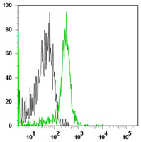
FITC anti-human CD1a

Human T lymphoblastic leukemia cell line Molt-4 stained with... -
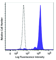
PE anti-human CD1a

Molt-4 (T leukemia cell line) stained with HI149 PE -
PE/Cyanine5 anti-human CD1a

Human T lymphoblastic leukemia cell line Molt-4 stained with... -
APC anti-human CD1a

Human T lymphoblastic leukemia cell line Molt-4 stained with... -
Purified anti-human CD1a

Human T lymphoblastic leukemia cell line Molt-4 stained with... -
Biotin anti-human CD1a

Human T lymphoblastic leukemia cell line Molt-4 stained with... -
Alexa Fluor® 488 anti-human CD1a

Human T lymphoblastic leukemia cell line Molt-4 stained with... -
Alexa Fluor® 647 anti-human CD1a

Human T lymphoblastic leukemia cell line Molt-4 stained with... -
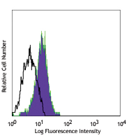
Pacific Blue™ anti-human CD1a

Human T leukemia cell line, Molt-4, stained with HI149 Pacif... -
Alexa Fluor® 700 anti-human CD1a

Human T lymphoblastic leukemia cell line Molt-4 stained with... -
Brilliant Violet 421™ anti-human CD1a

Human T leukemia cell line (MOLT-4) was stained with CD1a (c... -
PE/Cyanine7 anti-human CD1a

Human T lymphoblastic leukemia cell line Molt-4 stained with... -
APC/Cyanine7 anti-human CD1a

Human T-lymphoblastic leukemia cell line Molt-4 were stained... -
PerCP/Cyanine5.5 anti-human CD1a

Human T leukemia cell line (MOLT-4) was stained with CD1a (c... -
PE/Dazzle™ 594 anti-human CD1a

Human T leukemia cell line (MOLT-4) was stained with CD1a (c... -
TotalSeq™-A0402 anti-human CD1a
-
TotalSeq™-C0402 anti-human CD1a
-
Brilliant Violet 711™ anti-human CD1a

Human T leukemia cell line (MOLT-4) was stained with CD1a (c... -
APC/Fire™ 750 anti-human CD1a

Human T leukemia cell line (MOLT-4) was stained with CD1a (c... -
Brilliant Violet 605™ anti-human CD1a

Human T leukemia cell line (MOLT-4) was stained with CD1a (c... -
TotalSeq™-B0402 anti-human CD1a
-
TotalSeq™-D0402 anti-human CD1a


Follow Us